From Pathology Education Instructional Resource
Jump to navigation
Jump to search
Images
-
This gross photograph of lung illustrates multiple
abscesses throughout the lung of this patient.
-
This is a closer view of the same lung tissue showing the individual small lung
abscesses (arrows).
-
This is a low-power photomicrograph of lung with multiple focal lesions (1) throughout the tissue, some of which have a pale center indicating a loss of parenchymal tissue (2). This is typical of
abscess formation in the lung and represents a form of liquefaction necrosis. This
pneumonia is bronchopneumonia since the distribution is along the bronchi and the terminal airway distribution throughout the lung. Note that there are some areas of lung which appear relatively normal, having a pale-staining appearance. Other areas, such as in the lower central area (3), are much darker, indicating a heavy cellular
infiltrate.
-
This photomicrograph of the wall of an
abscess (1) illustrates liquefaction necrosis in the center of the
abscess (2). The remaining lung parenchyma (on the right side of the image) has extensive neutrophilic infiltration into the alveoli (3). This
abscess has destroyed a portion of the lung, but other areas of the lung where the normal structure has been retained could recover normal function.
-
This is a photomicrograph of lung tissue affected by bronchopneumonia. Note that the alveolar structure of this tissue, which is in the region of a terminal bronchiole (1), has been retained despite the massive infiltration of inflammatory cells. These inflammatory cells are primarily neutrophils.
-
This is a photomicrograph of another area in the lung showing a terminal bronchiole (1) in which the mucosal lining has been almost completely destroyed. There is extensive neutrophilic infiltration throughout this lung tissue.
-
This higher-power photomicrograph shows a central portion of an
abscess. Note the absence of any parenchymal lung tissue in this section due to extensive neutrophilic infiltration with liquefaction necrosis of the parenchymal tissue. Masses of leukocytes (primarily neutrophils), fluid ("liquor puris" which is serum, fibrin, etc.), and necrotic debris within an
abscess form what is referred to as "purulent material" -- or "pus" in lay terminology. The blue-staining mass in the center of this
abscess (arrow) represents colonies of bacteria.
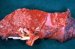 This gross photograph of lung illustrates multiple abscesses throughout the lung of this patient.
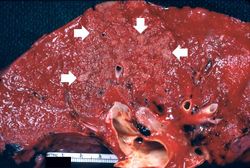 This is a closer view of the same lung tissue showing the individual small lung abscesses (arrows).
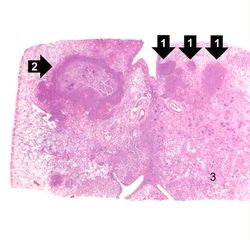 This is a low-power photomicrograph of lung with multiple focal lesions (1) throughout the tissue, some of which have a pale center indicating a loss of parenchymal tissue (2). This is typical of abscess formation in the lung and represents a form of liquefaction necrosis. This pneumonia is bronchopneumonia since the distribution is along the bronchi and the terminal airway distribution throughout the lung. Note that there are some areas of lung which appear relatively normal, having a pale-staining appearance. Other areas, such as in the lower central area (3), are much darker, indicating a heavy cellular infiltrate.
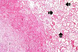 This photomicrograph of the wall of an abscess (1) illustrates liquefaction necrosis in the center of the abscess (2). The remaining lung parenchyma (on the right side of the image) has extensive neutrophilic infiltration into the alveoli (3). This abscess has destroyed a portion of the lung, but other areas of the lung where the normal structure has been retained could recover normal function.
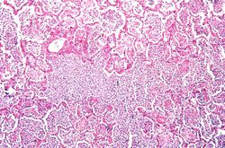 This is a photomicrograph of lung tissue affected by bronchopneumonia. Note that the alveolar structure of this tissue, which is in the region of a terminal bronchiole (1), has been retained despite the massive infiltration of inflammatory cells. These inflammatory cells are primarily neutrophils.
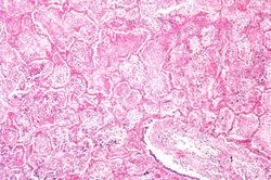 This is a photomicrograph of another area in the lung showing a terminal bronchiole (1) in which the mucosal lining has been almost completely destroyed. There is extensive neutrophilic infiltration throughout this lung tissue.
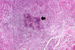 This higher-power photomicrograph shows a central portion of an abscess. Note the absence of any parenchymal lung tissue in this section due to extensive neutrophilic infiltration with liquefaction necrosis of the parenchymal tissue. Masses of leukocytes (primarily neutrophils), fluid ("liquor puris" which is serum, fibrin, etc.), and necrotic debris within an abscess form what is referred to as "purulent material" -- or "pus" in lay terminology. The blue-staining mass in the center of this abscess (arrow) represents colonies of bacteria.


