From Pathology Education Instructional Resource
Jump to navigation
Jump to search
Images
-
This is a head CT showing the two cysts (arrows).
-
This is a photograph of the cyst that was surgically removed. The cyst is filled with a clear fluid and contains a scolex.
-
This photograph of an autopsy specimen from another patient shows an adult tapeworm in the intestine. Note that the worm attaches to the luminal surface of the intestine via the scolex.
-
This is a photograph of the body of an adult tapeworm. Note the body segments.
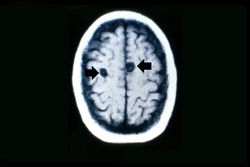 This is a head CT showing the two cysts (arrows).
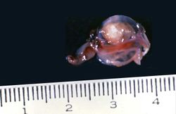 This is a photograph of the cyst that was surgically removed. The cyst is filled with a clear fluid and contains a scolex.
 This photograph of an autopsy specimen from another patient shows an adult tapeworm in the intestine. Note that the worm attaches to the luminal surface of the intestine via the scolex.
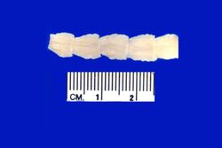 This is a photograph of the body of an adult tapeworm. Note the body segments.


