Clinical Summary[edit]
This 9-year-old child was admitted with headache, fever, and a morbilliform rash on the arms and legs. There was a history of a tick being removed from her back. By the time a biopsy was performed, the rash had become petechial. Antibiotics were given and the child recovered within one week.
Biopsy Findings[edit]
A skin biopsy of this patient's lesion was stained with hematoxylin and eosin. A different section was also stained with an immunoperoxidase technique using antibody against Rickettsia rickettsii. Organisms were demonstrated in the endothelial cells.
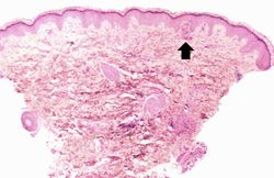
This is a low-power photomicrograph of the skin biopsy. Note areas of hemorrhage (arrow) in the dermis.
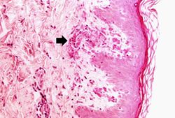
This is a higher-power photomicrograph demonstrating areas of hemorrhage immediately underneath the epidermis. Also note the cellularity and thrombosis of the small vessels in the dermis (arrow).
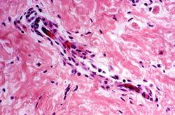
This is a photomicrograph of a small vessel in the dermis which is demonstrating mild vasculitis.
This is a higher-power photomicrograph of a dermal vessel with mild vasculitis.
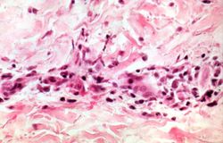
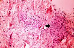
This is a photomicrograph of dermis with an area of more severe vasculitis (arrow).
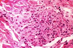
This is a high-power photomicrograph of severe vasculitis in the dermis.
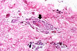
This is a high-power photomicrograph of a dermal vessel (arrow) which is exhibiting vasculitis and thrombosis.
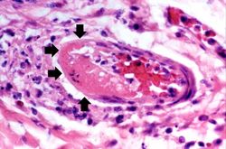
This is a high-power photomicrograph of a thrombosed vessel in the dermis. Note that the endothelial cells are missing along part of the circumference of the vessel (arrows)--this is where the main part of the thrombus has attached. Also note the inflammation surrounding the vessel.
Study Questions[edit]
Rickettsia rickettsii. Rodent and dog ticks transmit R. rickettsii to man. Rickettsiae enter the skin with the bite or with scratching of skin covered with insect feces.
An eschar develops at the site of the tick bite followed by a hemorrhagic rash that extends over the entire body, including the palms of the hands and soles of the feet. The vascular lesions that underlie the rash often lead to acute necrosis, fibrin extravasation, and thrombosis of the small blood vessels, including arterioles. In severe RMSF, foci of necrotic skin can form on the fingers, toes, elbows, ears, and scrotum.
Rickettsiae predominantly infect host endothelial and vascular smooth muscle cells causing a widespread vasculitis that may be complicated by thrombi and hemorrhages. Rickettsiae bind to cholesterol-containing receptors, are endocytosed into phagolysosomes, escape into the cytosol, and multiply until they burst the cells. Rickettsiae have an endotoxin but lack secreted toxins. In addition, R. rickettsii activate host kallikrein and kinins and cause local clotting.
Additional Resources[edit]
Reference[edit]
Journal Articles[edit]
