IPLab:Lab 1:Lung Abscess: Difference between revisions
| Line 7: | Line 7: | ||
File:IPLab1LungAbscess1.jpg|This is a gross photograph of the lungs from this case. Note the abscesses (arrows) especially in the lower lobes. The entire lung is consolidated. | File:IPLab1LungAbscess1.jpg|This is a gross photograph of the lungs from this case. Note the abscesses (arrows) especially in the lower lobes. The entire lung is consolidated. | ||
File:IPLab1LungAbscess2.jpg|This is a closer view of the lung from this case. In this section of upper lobe there are multiple areas of early abscess formation (arrows). Note the circumscribed whitish-tan lesions. These lesions are filled with white blood cells. | File:IPLab1LungAbscess2.jpg|This is a closer view of the lung from this case. In this section of upper lobe there are multiple areas of early abscess formation (arrows). Note the circumscribed whitish-tan lesions. These lesions are filled with white blood cells. | ||
File:IPLab1LungAbscess3.jpg|This low-power photomicrograph of lung from this case demonstrates one of the abscesses (arrows). Note that the material inside the abscess has been | File:IPLab1LungAbscess3.jpg|This low-power photomicrograph of lung from this case demonstrates one of the abscesses (arrows). Note that the material inside the abscess has been degraded and removed. Note the loose necrotic material inside the abscess. This material is made up of inflammatory cells (primarily dead white blood cells) and necrotic lung tissue. | ||
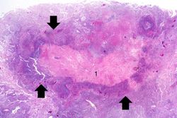
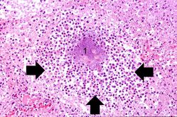
File:IPLab1LungAbscess5.jpg|This is a low-power photomicrograph of lung tissue containing a large abscess. The center of the abscess contains necrotic debris (1) and there is a rim of viable inflammatory cells (arrows) surrounding this abscess. | File:IPLab1LungAbscess5.jpg|This is a low-power photomicrograph of lung tissue containing a large abscess. The center of the abscess contains necrotic debris (1) and there is a rim of viable inflammatory cells (arrows) surrounding this abscess. | ||
File:IPLab1LungAbscess6.jpg|This high-power photomicrograph demonstrates a small abscess (arrow) with a necrotic center. | File:IPLab1LungAbscess6.jpg|This high-power photomicrograph demonstrates a small abscess (arrow) with a necrotic center. | ||
Revision as of 21:36, 27 June 2019
Clinical Summary
This 67-year-old male with advanced colon cancer underwent palliative surgery to remove a bowel obstruction. During the surgery the patient had several episodes of hypotension and after surgery he required ventilator support. Four days later the patient developed a fever and his white blood cell count was 15,256 cells/mm3. A chest x-ray demonstrated infiltrates in both lungs, which worsened over the next several days. His overall condition continued to deteriorate and he died 12 days after surgery. At autopsy, the lungs were markedly consolidated and had several focal abscesses that were 2 to 4 cm in diameter. Liquefied material poured out from these abscesses when the lungs were sliced.
Images
-
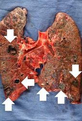 This is a gross photograph of the lungs from this case. Note the abscesses (arrows) especially in the lower lobes. The entire lung is consolidated.
 -
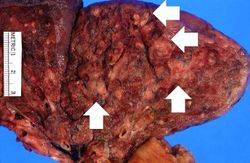 This is a closer view of the lung from this case. In this section of upper lobe there are multiple areas of early abscess formation (arrows). Note the circumscribed whitish-tan lesions. These lesions are filled with white blood cells.
 -
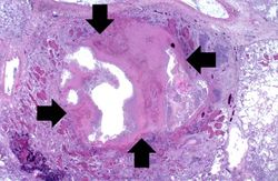 This low-power photomicrograph of lung from this case demonstrates one of the abscesses (arrows). Note that the material inside the abscess has been degraded and removed. Note the loose necrotic material inside the abscess. This material is made up of inflammatory cells (primarily dead white blood cells) and necrotic lung tissue.
 -
-
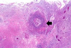 This high-power photomicrograph demonstrates a small abscess (arrow) with a necrotic center.
 -
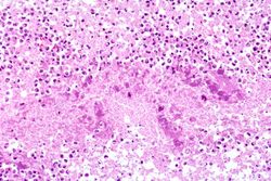 This high-power photomicrograph shows the center of an abscess containing neutrophils and necrotic debris.
 -


Virtual Microscopy
Lung Abscess
<peir-vm>IPLab1LungAbscess</peir-vm>
Normal Lung
<peir-vm>UAB-Histology-00107</peir-vm>
Study Questions
- <spoiler text="What factors predisposed this patient to pneumonia?">The patient had terminal cancer, had undergone a major surgery, and was on a ventilator. Any of these factors could have predisposed the patient to pneumonia. Also, being a patient in the hospital is itself a risk factor due to nosocomial infections.</spoiler>
- <spoiler text="What causes abscesses to liquefy?">The proteolytic enzymes released by white blood cells digest the tissue and cause it to liquefy. The presence of bacteria accentuates the inflammatory response and the bacteria themselves can also release enzymes that digest the tissue.</spoiler>
- <spoiler text="Had the patient lived, could these lesions have resolved?">If the patient had lived, the body could have eventually removed all of the necrotic and liquefied material. However, since there was loss of tissue and disruption of the basement membranes, many of the lesions would heal by scar tissue. So, the lung would never return to its original normal morphology and function.</spoiler>
Additional Resources
Reference
Journal Articles
- Brandenburg JA et al. Clinical presentation, processes and outcomes of care for patients with Pneumococcal pneumonia. J Gen Intern Med 2000 September; 15(9): 638–646.
Images
Related IPLab Cases
| |||||