IPLab:Lab 11:Cysticercosis: Difference between revisions
Jump to navigation
Jump to search
Seung Park (talk | contribs) No edit summary |
Seung Park (talk | contribs) No edit summary |
||
| Line 16: | Line 16: | ||
== Study Questions == | == Study Questions == | ||
* <spoiler text="How did this patient get this parasite?">Cysticercosis is caused by the cestode parasite, Taenia solium (pig tapeworm). Cysticercosis is contracted by ingesting eggs from the feces of a person infected with an adult Taenia solium. The eggs can survive for several weeks in soil but are also infectious as soon as passed. The larvae migrate through the intestinal wall and are disseminated via the blood stream and can encyst in the brain, heart, skeletal muscle, or skin.</spoiler> | * <spoiler text="How did this patient get this parasite?">Cysticercosis is caused by the cestode parasite, Taenia solium (pig tapeworm). Cysticercosis is contracted by ingesting eggs from the feces of a person infected with an adult Taenia solium. The eggs can survive for several weeks in soil but are also infectious as soon as passed. The larvae migrate through the intestinal wall and are disseminated via the blood stream and can encyst in the brain, heart, skeletal muscle, or skin.</spoiler> | ||
== Additional Resources == | |||
=== Reference === | |||
=== Journal Articles === | |||
=== Images === | |||
== Related IPLab Cases == | |||
{{IPLab 11}} | {{IPLab 11}} | ||
[[Category: IPLab:Lab 11]] | [[Category: IPLab:Lab 11]] | ||
Revision as of 23:25, 22 August 2013
Clinical Summary
This 29-year-old woman was admitted to the hospital because of repeated tonic-clonic seizures. The patient was a tour guide leading groups of tourist to Tibet for two-month walking/camping tours in the Himalayas. Her seizures were easily controlled by intravenous administration of phenytoin. The WBC count was 13,000, with 5% eosinophils; the erythrocyte sedimentation rate was 19. A cranial CT performed with and without contrast revealed two ring-enhancing lesions. The patient underwent a craniotomy and excisional biopsy.
Surgical Specimen
Histopathologic exam revealed a capsule of dense connective tissue surrounding a cavity that contained a partially degenerated scolex of Taenia solium.
Images
-
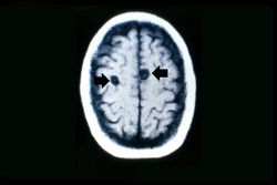 This is a head CT showing the two cysts (arrows).
 -
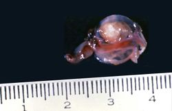 This is a photograph of the cyst that was surgically removed. The cyst is filled with a clear fluid and contains a scolex.
 -
 This photograph of an autopsy specimen from another patient shows an adult tapeworm in the intestine. Note that the worm attaches to the luminal surface of the intestine via the scolex.
 -
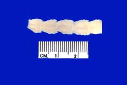 This is a photograph of the body of an adult tapeworm. Note the body segments.


Study Questions
Additional Resources
Reference
Journal Articles
Images
Related IPLab Cases
| |||||