IPLab:Lab 3:Bronchopneumonia: Difference between revisions
Jump to navigation
Jump to search
Seung Park (talk | contribs) No edit summary |
Seung Park (talk | contribs) No edit summary |
||
| Line 16: | Line 16: | ||
File:IPLab3Bronchopneumonia7.jpg|This higher-power photomicrograph shows a central portion of an abscess. Note the absence of any parenchymal lung tissue in this section due to extensive neutrophilic infiltration with liquefaction necrosis of the parenchymal tissue. Masses of leukocytes (primarily neutrophils), fluid ("liquor puris" which is serum, fibrin, etc.), and necrotic debris within an abscess form what is referred to as "purulent material" -- or "pus" in lay terminology. The blue-staining mass in the center of this abscess (arrow) represents colonies of bacteria. | File:IPLab3Bronchopneumonia7.jpg|This higher-power photomicrograph shows a central portion of an abscess. Note the absence of any parenchymal lung tissue in this section due to extensive neutrophilic infiltration with liquefaction necrosis of the parenchymal tissue. Masses of leukocytes (primarily neutrophils), fluid ("liquor puris" which is serum, fibrin, etc.), and necrotic debris within an abscess form what is referred to as "purulent material" -- or "pus" in lay terminology. The blue-staining mass in the center of this abscess (arrow) represents colonies of bacteria. | ||
</gallery> | </gallery> | ||
Study Questions | |||
* <spoiler text="What does it mean to have a shift to the left?">A "shift to the left" indicates an increased ratio of immature PMNs ("bands") to mature PMNs ("segs") within the peripheral circulation.</spoiler> | |||
* <spoiler text="Is there destruction of lung parenchyma in bronchopneumonia?">Yes, there is usually destruction of alveoli and, in this case, there are abscesses containing bacteria.</spoiler> | |||
* <spoiler text="How would this lung have healed if the patient had survived?">Scar tissue would form in the areas of tissue destruction (abscesses).</spoiler> | |||
{{IPLab 3}} | {{IPLab 3}} | ||
Revision as of 23:51, 18 August 2013
Clinical Summary
This 15-year-old black female sustained third degree burns involving approximately 85% of the body surface. On admission to the hospital, the patient was taken to the operating room where a tracheotomy was performed and her burned body surface was debrided. After a few days of hospitalization, the peripheral white blood count was 41,000 cells/mm³ with a shift to the left. In spite of intensive therapy, which included administration of fluids and antibiotics, the patient expired on the sixth hospital day.
Autopsy Findings
Each lung weighed approximately 630 grams and was studded with gray nodules which ranged in size from 3 to 6 mm in diameter. In several areas, these coalesced to form nodules having necrotic-appearing centers.
Images
-
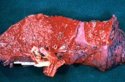 This gross photograph of lung illustrates multiple abscesses throughout the lung of this patient.
 -
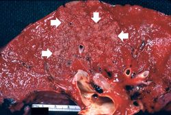 This is a closer view of the same lung tissue showing the individual small lung abscesses (arrows).
 -
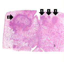 This is a low-power photomicrograph of lung with multiple focal lesions (1) throughout the tissue, some of which have a pale center indicating a loss of parenchymal tissue (2). This is typical of abscess formation in the lung and represents a form of liquefaction necrosis. This pneumonia is bronchopneumonia since the distribution is along the bronchi and the terminal airway distribution throughout the lung. Note that there are some areas of lung which appear relatively normal, having a pale-staining appearance. Other areas, such as in the lower central area (3), are much darker, indicating a heavy cellular infiltrate.
 -
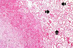 This photomicrograph of the wall of an abscess (1) illustrates liquefaction necrosis in the center of the abscess (2). The remaining lung parenchyma (on the right side of the image) has extensive neutrophilic infiltration into the alveoli (3). This abscess has destroyed a portion of the lung, but other areas of the lung where the normal structure has been retained could recover normal function.
 -
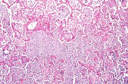 This is a photomicrograph of lung tissue affected by bronchopneumonia. Note that the alveolar structure of this tissue, which is in the region of a terminal bronchiole (1), has been retained despite the massive infiltration of inflammatory cells. These inflammatory cells are primarily neutrophils.
 -
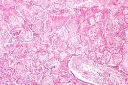 This is a photomicrograph of another area in the lung showing a terminal bronchiole (1) in which the mucosal lining has been almost completely destroyed. There is extensive neutrophilic infiltration throughout this lung tissue.
 -
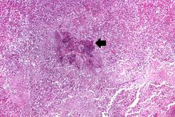 This higher-power photomicrograph shows a central portion of an abscess. Note the absence of any parenchymal lung tissue in this section due to extensive neutrophilic infiltration with liquefaction necrosis of the parenchymal tissue. Masses of leukocytes (primarily neutrophils), fluid ("liquor puris" which is serum, fibrin, etc.), and necrotic debris within an abscess form what is referred to as "purulent material" -- or "pus" in lay terminology. The blue-staining mass in the center of this abscess (arrow) represents colonies of bacteria.


Study Questions