IPLab:Lab 3:Brain Infarction: Difference between revisions
| Line 11: | Line 11: | ||
File:IPLab3BrainInfarction4b.JPG|This photomicrograph shows the area of healed/healing infarction (I) and more normal brain parenchyma (B). | File:IPLab3BrainInfarction4b.JPG|This photomicrograph shows the area of healed/healing infarction (I) and more normal brain parenchyma (B). | ||
File:IPLab3BrainInfarction5b.JPG|This is a higher-power photomicrograph of the previous image showing the area of healed/healing infarction (I) and more normal brain parenchyma (B). In the infarcted area contains numerous microglia (brain macrophages) that have phagocytosed dead brain cell debris that are called "gitter cells". In the more normal brain tissue there are several enlarged astrocytes. These activated astrocytes are often seen in areas of healing and they are called "gemistocytic astrocytes". | File:IPLab3BrainInfarction5b.JPG|This is a higher-power photomicrograph of the previous image showing the area of healed/healing infarction (I) and more normal brain parenchyma (B). In the infarcted area contains numerous microglia (brain macrophages) that have phagocytosed dead brain cell debris that are called "gitter cells". In the more normal brain tissue there are several enlarged astrocytes. These activated astrocytes are often seen in areas of healing and they are called "gemistocytic astrocytes". | ||
File:IPLab3BrainInfarction6b.JPG|This is a photomicrograph of | File:IPLab3BrainInfarction6b.JPG|This is a higher-power photomicrograph of the brain tissue adjacent to the infarct. In this image there are numerous enlarged activated astrocytes that are often seen in areas of brain healing - gemistocytic astrocytes (arrows). | ||
File:IPLab3BrainInfarction7b.JPG|This is a photomicrograph of | File:IPLab3BrainInfarction7b.JPG|This is a higher-power photomicrograph of infarcted brain tissue with numerous macrophages (microglia) that are full of brain tissue - gitter cells (arrows). | ||
File:IPLab3BrainInfarction10.jpg|This is a gross photograph of a brain from another patient with an old healed infarct. Note the meninges overlying the infarcted region (arrow). | File:IPLab3BrainInfarction10.jpg|This is a gross photograph of a brain from another patient with an old healed infarct. Note the meninges overlying the infarcted region (arrow). | ||
File:IPLab3BrainInfarction11.jpg|This is a closer view of the brain demonstrating an old healed infarct with the meninges containing blood vessels (arrow) overlying the infarcted region. | File:IPLab3BrainInfarction11.jpg|This is a closer view of the brain demonstrating an old healed infarct with the meninges containing blood vessels (arrow) overlying the infarcted region. | ||
Latest revision as of 17:54, 23 June 2020
Clinical Summary
Eight years prior to his demise, this 48-year-old black male had a dissecting aortic aneurysm. The first portion of the aortic arch was dilated and he had associated aortic valve insufficiency. The defect was repaired and a mechanical prosthetic aortic valve was implanted. One year prior to his terminal admission, the patient experienced a catastrophic cerebrovascular accident (CVA) with left hemiparesis and obtundation. The patient experienced severe neurologic sequelae from this CVA and was eventually transferred to a nursing home where he was bedbound and required an indwelling urinary catheter. Four months later the patient developed a urinary tract infection and was transferred to the hospital. The patient's condition deteriorated with a urinary tract infection and pneumonia and he died of sepsis.
Autopsy showed the expected findings in the aorta and in a 760-gram heart. The mechanical valve showed some old thrombotic material adherent to the valve which was probably the source of the emboli which caused the patient's CVA. The patient's 1250-gram brain showed extensive old infarctions in at least three different areas, with the largest (12.5 x 5.5 cm) in the distribution of the right middle cerebral artery.
Images
-
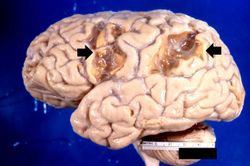 This is a gross photograph of the brain which contains two areas of infarction (arrows).
 -
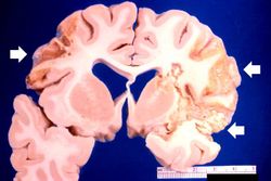 This is a gross photograph of a cross-section of brain demonstrating the areas of infarction (arrows).
 -
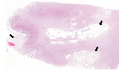 This is a low-power photomicrograph of this brain with several areas of infarcted tissue. Note the pale areas which correspond to areas with loss of brain parenchyma (arrows).
 -
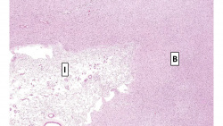 This photomicrograph shows the area of healed/healing infarction (I) and more normal brain parenchyma (B).
 -
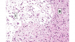 This is a higher-power photomicrograph of the previous image showing the area of healed/healing infarction (I) and more normal brain parenchyma (B). In the infarcted area contains numerous microglia (brain macrophages) that have phagocytosed dead brain cell debris that are called "gitter cells". In the more normal brain tissue there are several enlarged astrocytes. These activated astrocytes are often seen in areas of healing and they are called "gemistocytic astrocytes".
 -
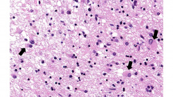 This is a higher-power photomicrograph of the brain tissue adjacent to the infarct. In this image there are numerous enlarged activated astrocytes that are often seen in areas of brain healing - gemistocytic astrocytes (arrows).
 -
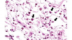 This is a higher-power photomicrograph of infarcted brain tissue with numerous macrophages (microglia) that are full of brain tissue - gitter cells (arrows).
 -
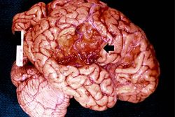 This is a gross photograph of a brain from another patient with an old healed infarct. Note the meninges overlying the infarcted region (arrow).
 -
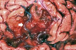 This is a closer view of the brain demonstrating an old healed infarct with the meninges containing blood vessels (arrow) overlying the infarcted region.


Study Questions
Additional Resources
Reference
- eMedicine Medical Library: Ischemic Stroke
- eMedicine Medical Library: Acute Aortic Dissection
- eMedicine Medical Library: Prosthetic Heart Valves
- Merck Manual: Overview of Stroke (CVA)
- Merck Manual: Thrombotic Disorders
- Merck Manual: Aortic Dissection
Journal Articles
- Love S, Barber R, Wilcock GK. Neuronal death in brain infarcts in man. Neuropathol Appl Neurobiol 2000 Feb;26(1):55-66.