IPLab:Lab 1:Prostate: Difference between revisions
Jump to navigation
Jump to search
| Line 1: | Line 1: | ||
== Clinical Summary == | == Clinical Summary == | ||
This 68-year-old man with a 7-year history of prostatic carcinoma developed metastases to the bones of the pelvis and lumbar spine which produced significant pain and discomfort. He was treated with androgen deprivation therapy to slow the growth of the tumor and decrease the size of the tumor metastases in order to help relieve his bone pain. Two months after starting the androgen deprivation therapy, the patient experienced an acute myocardial infarction and died suddenly. At autopsy there was evidence of cancer in the peripheral region of the prostate gland and there were metastases present in the pelvis and lumbar spine. The patient also had moderate testicular atrophy. | This 68-year-old man with a 7-year history of prostatic carcinoma developed metastases to the bones of the pelvis and lumbar spine which produced significant pain and discomfort. He was treated with androgen deprivation therapy to slow the growth of the tumor and decrease the size of the tumor metastases in order to help relieve his bone pain. Two months after starting the androgen deprivation therapy, the patient experienced an acute myocardial infarction and died suddenly. At autopsy there was evidence of cancer in the peripheral region of the prostate gland and there were metastases present in the pelvis and lumbar spine. The patient also had moderate testicular atrophy. | ||
== Images == | == Images == | ||
Latest revision as of 22:10, 27 June 2019
Clinical Summary
This 68-year-old man with a 7-year history of prostatic carcinoma developed metastases to the bones of the pelvis and lumbar spine which produced significant pain and discomfort. He was treated with androgen deprivation therapy to slow the growth of the tumor and decrease the size of the tumor metastases in order to help relieve his bone pain. Two months after starting the androgen deprivation therapy, the patient experienced an acute myocardial infarction and died suddenly. At autopsy there was evidence of cancer in the peripheral region of the prostate gland and there were metastases present in the pelvis and lumbar spine. The patient also had moderate testicular atrophy.
Images
-
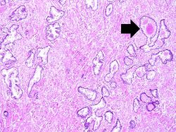 This is a low-power photomicrograph of a normal area from this patient's prostate. Note the corpora amylacea (arrow).
 -
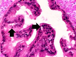 This high-power photomicrograph of the prostatic epithelium shows occasional cells with pyknotic fragmented nuclei (arrows).
 -
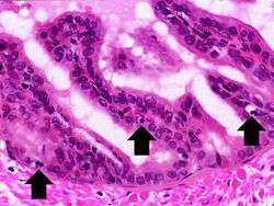 Another high-power photomicrograph of the prostatic epithelium shows cells with pyknotic and fragmented nuclei (arrows). Note that the cytoplasm is condensed and hypereosinophilic.
 -
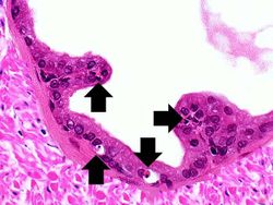 Still another high-power photomicrograph of the prostatic epithelium demonstrates cells with pyknotic and fragmented nuclei (arrows). Again note the condensed and hypereosinophilic cytoplasm.
 -
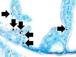 This photomicrograph of prostatic epithelium demonstrates an in situ immunohistochemical technique that is used to identify the DNA fragments characteristic of apoptotic nuclei. This technique, terminal deoxynucleotidyl transferase-mediated dUTP-biotin nick end-labeling (TUNEL) is used to identify apoptotic cells (arrows) in histology sections.
 -
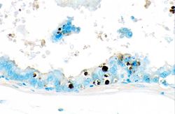 This is a higher-power photomicrograph of prostatic epithelium with the TUNEL staining. Note the apoptotic cells (brown nuclei) in the epithelium as well as those floating freely.


Study Questions
- <spoiler text="What is the cause of apoptosis in this case?">The diethylstilbesterol blocks the normal trophic hormone response that controls prostate growth. With loss of trophic influence the prostatic epithelial cells go through apoptosis and this results in atrophy of the gland. This would be an example of pathologic atrophy of a hormone-dependent tissue.</spoiler>
- <spoiler text="How is apoptosis different from necrosis?">Apoptosis is the result of a gene program that leads to self-destruction of individual cells. The apoptotic cells shrink, there is condensation of the chromatin, formation of cytoplasmic blebs, and fragmentation of the cell to form apoptotic bodies. Apoptotic bodies are membrane-bound fragments of cytoplasmic organelles with or without fragments of chromatin. A hallmark of apoptosis distinguishing it from necrosis is the fact that there is no inflammation associated with apoptotic cell death as compared to other types of cell death (e.g., necrosis).</spoiler>
Additional Resources
Reference
- eMedicine Medical Library: Metastatic and Advanced Prostate Cancer
- Merck Manual: Prostate Cancer
- National Cancer Institute: Prostate Cancer
- British Columbia Cancer Agency: Prostate Cancer
Journal Articles
- Denmeade SR, Isaacs JT. Programmed cell death (apoptosis) and cancer chemotherapy. Cancer Control 1996 Jul;3(4):303-309.
- Brookes PS, Salinas EP, Darley-Usmar K, Eiserich JP, Freeman BA, Darley-Usmar VM, Anderson PG. Concentration-dependent effects of nitric oxide on mitochondrial permeability transition and cytochrome c release. J Biol Chem 2000 Jul 7;275(27):20474-9.
Images
| |||||