IPLab:Lab 1:Tuberculosis: Difference between revisions
| Line 14: | Line 14: | ||
File:IPLab1Tuberculosis6.jpg|This photomicrograph shows a single nodule with an amorphous eosinophilic center and accumulations of cells around the outer edge. This is typical of a granuloma associated with tuberculosis in which there is a necrotic center (1) and a rim of lymphocytes, macrophages, and occasional multinucleated giant cells around the periphery. | File:IPLab1Tuberculosis6.jpg|This photomicrograph shows a single nodule with an amorphous eosinophilic center and accumulations of cells around the outer edge. This is typical of a granuloma associated with tuberculosis in which there is a necrotic center (1) and a rim of lymphocytes, macrophages, and occasional multinucleated giant cells around the periphery. | ||
File:IPLab1Tuberculosis7.jpg|This is a higher-power view of the granuloma with the amorphous eosinophilic material representing caseation necrosis (1), giant cells near the center (2), and inflammatory cells around the periphery. | File:IPLab1Tuberculosis7.jpg|This is a higher-power view of the granuloma with the amorphous eosinophilic material representing caseation necrosis (1), giant cells near the center (2), and inflammatory cells around the periphery. | ||
File:IPLab1Tuberculosis8.jpg|This is a high-power photomicrograph of | File:IPLab1Tuberculosis8.jpg|This is a high-power photomicrograph of Langhans-type multinucleated giant cells (arrows) that are characteristic of tuberculous granulomas. Note the ring of the nuclei in these giant cells. | ||
</gallery> | </gallery> | ||
Revision as of 13:48, 31 October 2016
Clinical Summary
This 70-year-old man was admitted to the hospital with a history of upper abdominal pain, anorexia, nausea, and general malaise, all of approximately three weeks' duration. His hospital stay was characterized by fever and severe respiratory distress. There were multiple densities in the patient's chest x-ray consistent with pneumonia and examination of a stained sputum specimen showed acid fast bacilli. Despite intensive therapy, the patient progressively deteriorated and died 14 days after admission.
Autopsy Findings
It was determined at autopsy that the patient suffered from pulmonary tuberculosis with widespread dissemination throughout the body. The left lung weighed 620 grams and the right lung 1230 grams. These were characterized by marked pulmonary congestion and pulmonary edema. In addition, multiple gray-white nodules ranging from pinpoint size up to 1 cm were diffusely distributed throughout the lung parenchyma.
Images
-
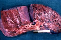 This is a gross photograph of a cut section of lung from this patient with disseminated tuberculosis. The numerous small white nodules scattered throughout this lung tissue represent individual tuberculosis granulomas. In addition, note the dark areas throughout the lung which represent deposits of anthracotic pigment.
 -
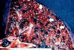 This is a closer view of the same section of lung containing multiple white granulomas which are now more easily identified (arrows). These lesions are referred to as miliary tuberculosis. Dark areas of anthracosis are also prominent in this lung.
 -
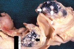 This gross photograph shows hilar lymph nodes from another patient with disseminated tuberculosis. The white, cheesy-appearing nodules (arrows) in the lymph nodes give rise to the descriptive terminology of caseous necrosis. The black pigment in the lymph nodes is anthracotic pigment that has drained from the lungs.
 -
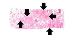 This is a low-power photomicrograph of a histology section from the lung of this patient with a chronic history of respiratory disease. Note the multiple eosinophilic nodules (arrows) seen at low power in this section. Other areas of the lung are relatively normal and several bronchi and large vessels can be seen at this low power. The pleural surface of the lung is at the left and the remaining edges are cut edges of the tissue block.
 -
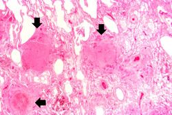 This higher-power photomicrograph of the eosinophilic nodules (arrows) illustrates their discreet nature and the surrounding inflammatory response in the remaining normal lung tissue.
 -
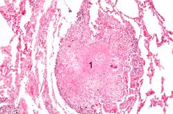 This photomicrograph shows a single nodule with an amorphous eosinophilic center and accumulations of cells around the outer edge. This is typical of a granuloma associated with tuberculosis in which there is a necrotic center (1) and a rim of lymphocytes, macrophages, and occasional multinucleated giant cells around the periphery.
 -
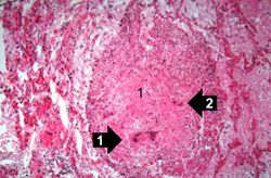 This is a higher-power view of the granuloma with the amorphous eosinophilic material representing caseation necrosis (1), giant cells near the center (2), and inflammatory cells around the periphery.
 -
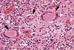 This is a high-power photomicrograph of Langhans-type multinucleated giant cells (arrows) that are characteristic of tuberculous granulomas. Note the ring of the nuclei in these giant cells.


Virtual Microscopy
Lung: Tuberculosis
Normal Lung
Study Questions
Additional Resources
Reference
- eMedicine Medical Library: Tuberculosis
- Merck Manual: Tuberculosis
- Centers for Disease Control: Tuberculosis
Journal Articles
- Lee MP, Chan JW, Ng KK, Li PC. Clinical manifestations of tuberculosis in HIV-infected patients. Respirology 2000 Dec;5(4):423-6.
Images
Related IPLab Cases
| |||||