IPLab:Lab 3:Foreign Body Granuloma: Difference between revisions
Jump to navigation
Jump to search
Seung Park (talk | contribs) |
Seung Park (talk | contribs) No edit summary |
||
| Line 10: | Line 10: | ||
File:IPLab3ForeignBodyGranuloma5.jpg|This is a fully-polarized view of lung showing numerous birefringent particles. | File:IPLab3ForeignBodyGranuloma5.jpg|This is a fully-polarized view of lung showing numerous birefringent particles. | ||
</gallery> | </gallery> | ||
== Virtual Microscopy == | |||
<peir-vm>IPLab3ForeignBodyGranuloma</peir-vm> | |||
== Study Questions == | == Study Questions == | ||
* <spoiler text="Compare and contrast the tissue reactions to carbon (anthracosis), talc, and silica. What factors are important in the pathogenesis of tissue injury due to these foreign materials?">Carbon is relatively inert and leads to little if any tissue damage. | * <spoiler text="Compare and contrast the tissue reactions to carbon (anthracosis), talc, and silica. What factors are important in the pathogenesis of tissue injury due to these foreign materials?">Carbon is relatively inert and leads to little if any tissue damage. | ||
Revision as of 11:07, 3 January 2014
Clinical Summary
This 21-year-old black male suffered a gunshot wound to his right chest. A thoracotomy revealed multiple bleeding points in the middle and lower lobe of the right lung and significant hemorrhage from an intercostal artery. A portion of the right middle lobe was removed. The patient's post-operative course was complicated by Gram-negative pneumonia and significant neurologic dysfunction, apparently the result of emboli from iliofemoral venous thrombosis. All were treated without sequelae.
Images
-
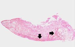 This is a low-power photomicrograph of lung and pleura. There is some hemorrhage in this tissue (arrows), probably the result of surgery or the gunshot wounds.
 -
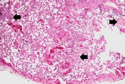 This higher-power photomicrograph demonstrates the small focal cellular lesions (arrows) found throughout the parenchyma of the lung.
 -
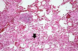 A higher-power photomicrograph of these granulomas reveals that they surround blood vessels (note the red blood cells within the lumen) (arrow).
 -
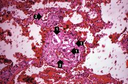 This is a photomicrograph of lung taken under light that is partially polarized to demonstrate the birefringent particles within the granulomas (1). Also, at this magnification one can better appreciate that these granulomas are adjacent to blood vessels (2).
 -
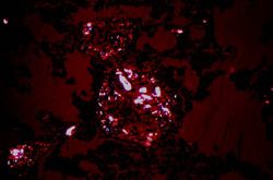 This is a fully-polarized view of lung showing numerous birefringent particles.


Virtual Microscopy
Study Questions
Additional Resources
Reference
- eMedicine Medical Library: Opioid Abuse
- Merck Manual: Opioids: Drug Use and Dependence
- Merck Manual: Occupational Asthma
Journal Articles
- Nan DN, Fernández-Ayala M, Iglesias L, García-Palomo D, Parra JA, Fariñas MC. Talc granulomatosis. A differential diagnosis of interstitial lung disease in HIV patients. Chest 2000 Jul;118(1):258-60.