IPLab:Lab 3:Acute Appendicitis: Difference between revisions
Jump to navigation
Jump to search
Seung Park (talk | contribs) |
Seung Park (talk | contribs) No edit summary |
||
| Line 16: | Line 16: | ||
File:IPLab3AcuteAppendicitis8.jpg|This is a gross photograph of another example of peritonitis. Again note the fibrinosuppurative exudate covering the abdominal organs (arrows). | File:IPLab3AcuteAppendicitis8.jpg|This is a gross photograph of another example of peritonitis. Again note the fibrinosuppurative exudate covering the abdominal organs (arrows). | ||
</gallery> | </gallery> | ||
== Virtual Microscopy == | |||
<peir-vm>IPLab3AcuteAppendicitis</peir-vm> | |||
== Study Questions == | == Study Questions == | ||
Revision as of 11:04, 3 January 2014
Clinical Summary
This 11-year-old male was admitted to the hospital with a complaint of 8 hours of severe pain in the right lower quadrant of the abdomen accompanied by nausea, vomiting, and diarrhea. Additional features on admission included a temperature of 101° F and a leukocytosis of 21,200 cells/mm³ with a shift to the left. An exploratory laparotomy revealed an inflamed appendix, retrocecal in location, which was adherent to the wall of the colon.
Autopsy Findings
The serosal surface of the appendix was covered with friable granular material. The lumen was dilated and contained a purulent exudate as well as a fecalith. The wall measured up to 0.4 cm in thickness.
Images
-
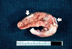 This is a gross photograph of the appendix which was removed from this patient with acute appendicitis. Note the rough, shaggy material (arrows) on the surface due to deposition of fibrin and inflammatory cells.
 -
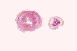 This is a low-power photomicrograph of a normal appendix on the right and an appendix with acute inflammatory response on the left. Note the abundant blue-stained lymphoid tissue beneath the mucosal layer and the absence of blue-staining cells in the submucosal layer of the normal appendix. Compare this with the extensive distribution of cells throughout the wall of the appendix with acute appendicitis. The blue color is due to the presence of many inflammatory cells, although at this low power these individual cells cannot be specifically identified.
 -
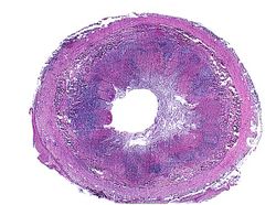 This is a photomicrograph of an appendix exhibiting acute inflammation. Note that there are only remnants of mucosal tissue identifiable along the luminal border of this specimen. There is an extensive infiltration of leukocytes in this tissue which cannot be specifically identified at this low power. Note that the surface is very roughened and has deposits of fibrin.
 -
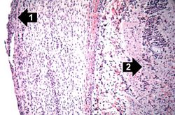 This is a photomicrograph of the serosal surface of the appendix on the left (1) and the submucosal tissue in the center (2) with remnants of a lymphoid nodule. Surrounding this lymphoid nodule are masses of leukocytes which should not be present in a normal appendix.
 -
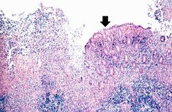 This photomicrograph of the mucosal surface shows a small area with normal mucosal epithelium (arrow). This area is surrounded by areas of ulceration with an inflammatory infiltrate of lymphocytes and neutrophils.
 -
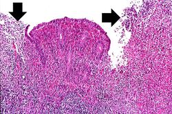 This higher-power photomicrograph of the mucosal surface shows the loss of normal mucosal epithelium (arrows) and the inflammatory infiltrate. The principal inflammatory cell in this case of acute appendicitis is the neutrophil.
 -
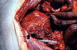 This is a gross photograph of the open abdominal cavity of a patient with acute appendicitis. In this patient, there had been rupture of the appendix with spillage of intestinal contents into the abdominal cavity. This spillage resulted in an acute abdomen with widespread inflammation throughout the abdominal cavity. Note the roughened surface of the mesenteric tissue (arrow) due to deposition of fibrin over much of the surface.
 -
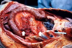 This is a gross photograph of another example of peritonitis. Again note the fibrinosuppurative exudate covering the abdominal organs (arrows).


Virtual Microscopy
Study Questions
Additional Resources
Reference
- eMedicine Medical Library: Appendicitis
- Merck Manual: Introduction to Acute Abdomen and Surgical Gastroenterology
- Merck Manual: Appendicitis
Journal Articles
- Helmer KS, Robinson EK, Lally KP, Vasquez JC, Kwong KL, Liu TH, Mercer DW. Standardized patient care guidelines reduce infectious morbidity in appendectomy patients. Am J Surg 2002 Jun;183(6):608-13.