IPLab:Lab 4:Mural Thrombus: Difference between revisions
Seung Park (talk | contribs) |
Seung Park (talk | contribs) |
||
| Line 37: | Line 37: | ||
=== Images === | === Images === | ||
* [ | * [{{SERVER}}/library/index.php?/tags/424-mural_thrombus PEIR Digital Library: Mural Thrombus Images] | ||
* [http://library.med.utah.edu/WebPath/CVHTML/CVIDX.html#3 WebPath: Myocardial Infarction] | * [http://library.med.utah.edu/WebPath/CVHTML/CVIDX.html#3 WebPath: Myocardial Infarction] | ||
* [http://library.med.utah.edu/WebPath/CVHTML/CVIDX.html#2 WebPath: Atherosclerotic Cardiovascular Disease] | * [http://library.med.utah.edu/WebPath/CVHTML/CVIDX.html#2 WebPath: Atherosclerotic Cardiovascular Disease] | ||
Revision as of 20:30, 29 August 2013
Clinical Summary
This 67-year-old female was transferred to the hospital from a nursing home in a comatose state. Physical findings on examination were compatible with brain stem infarction. On the fourth hospital day, an electrocardiogram revealed changes compatible with anterior myocardial infarction. The patient remained comatose with quadriplegia and expired on the 16th hospital day.
Autopsy Findings
Examination of the brain revealed extensive infarction involving the midbrain and cerebellum with complete occlusion of the upper one-half of the basilar artery; there was also extensive coronary artery atherosclerosis. A large aneurysm of the left ventricle was present; this was filled with mural thrombus. Extensive infarction of the lateral and posterior portions of the left ventricular wall toward the base of the heart was also found.
Images
-
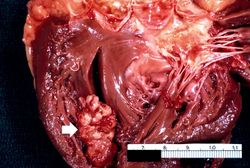 This is a gross photograph of the heart from this case demonstrating the well-formed thrombus (arrow) tightly attached to the myocardium near the apex of the left ventricle.
 -
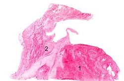 This is a low-power photomicrograph of the thrombus (1) attached to the myocardium (2).
 -
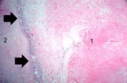 This higher-power photomicrograph shows the border between the thrombus on the right (1) and the endocardium on the left (2). There is a line of inflammatory cells at this interface (arrow).
 -
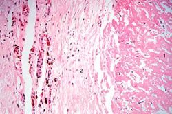 This is a high-power photomicrograph of the border zone between the thrombus (1) and the endocardium (2). In this region there is less inflammation at the border zone.
 -
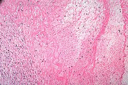 This photomicrograph illustrates the layered effect of the thrombus.
 -
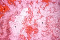 This is a higher-power photomicrograph of the thrombus. Note the pale regions which contain primarily platelets (degranulated platelets) with some fibrin (1), and the red areas which contain RBCs, some leukocytes, and fibrin(2).
 -
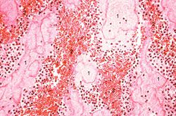 This high-power photomicrograph of thrombus demonstrates more clearly the components of the layers--the pale regions which contain primarily platelets (degranulated platelets) with some fibrin (1), and the red areas which contain RBCs, some leukocytes, and fibrin (2).


Study Questions
Additional Resources
Reference
- eMedicine Medical Library: Noncoronary Atherosclerosis
- eMedicine Medical Library: Myocardial Infarction
- Merck Manual: Thrombotic Disorders
- Merck Manual: Acute Coronary Syndromes (ACS)
Journal Articles
- Diop D, Aghababian RV. Definition, classification, and pathophysiology of acute coronary ischemic syndromes. Emerg Med Clin North Am 2001 May;19(2):259-67.
Images
- PEIR Digital Library: Mural Thrombus Images
- WebPath: Myocardial Infarction
- WebPath: Atherosclerotic Cardiovascular Disease
Related IPLab Cases
- Lab 1: Heart: Myocardial Infarction (Coagulative Necrosis)
- Lab 3: Heart: Acute Myocardial Infarction
- Lab 3: Heart: Healed Myocardial Infarction
- Lab 4: Coronary Artery: Thrombosis
- Lab 4: Lung: Pulmonary Congestion and Edema
| |||||