IPLab:Lab 9:ARF: Difference between revisions
Jump to navigation
Jump to search
Seung Park (talk | contribs) No edit summary |
Seung Park (talk | contribs) No edit summary |
||
| Line 21: | Line 21: | ||
This patient has acute ramifications of rheumatic fever (acute valve lesions and Aschoff bodies) but there are also a number of old lesions in the heart suggesting that this patient has had numerous bouts of rheumatic fever over the course of many years.</spoiler> | This patient has acute ramifications of rheumatic fever (acute valve lesions and Aschoff bodies) but there are also a number of old lesions in the heart suggesting that this patient has had numerous bouts of rheumatic fever over the course of many years.</spoiler> | ||
== Additional Resources == | |||
=== Reference === | |||
=== Journal Articles === | |||
=== Images === | |||
== Related IPLab Cases == | |||
{{IPLab 9}} | {{IPLab 9}} | ||
[[Category: IPLab:Lab 9]] | [[Category: IPLab:Lab 9]] | ||
Revision as of 23:23, 22 August 2013
Clinical Summary
This 21-year-old black male with sickle cell anemia had recurrent attacks of acute rheumatic fever beginning at age 14. Mitral insufficiency and stenosis were present by age 16. On prophylactic antibiotics, the patient had no evidence of recurrence until three weeks before his final admission, when an upper respiratory infection developed. A few weeks later he developed acute migratory polyarthritis. This was associated with rapid deterioration of cardiac function and death.
Autopsy Findings
At autopsy, the heart was enlarged--weighing 675 grams--especially the left atrium. Both the aortic and mitral valves showed fibrosis as well as the fresh, tiny verrucae characteristic of acute rheumatic fever.
Images
-
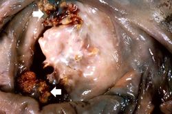 This is a gross photograph of mitral valve demonstrating marked thickening and fibrosis of the valve leaflet. There are also numerous foci of fibrinoid necrosis within the cusps and friable vegetations (verrucae) along the lines of closure (arrows). These irregular, warty projections are found at sites of erosion on the inflamed endocardial surface. The verrucae probably result from the precipitation of fibrin where the leaflets impinge on each other.
 -
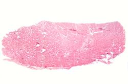 This is a low-power photomicrograph of heart tissue. Little can be seen at this magnification, except that the tissue looks relatively normal.
 -
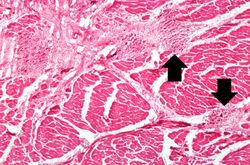 This is a higher-power photomicrograph of myocardium showing cellular accumulations--Aschoff bodies (arrows)--within the interstitium of the myocardium. These are found especially around blood vessels.
 -
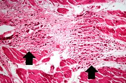 This is a higher-power photomicrograph of myocardium containing Aschoff bodies (arrows) within the interstitium.
 -
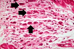 This high-power photomicrograph of myocardium shows the cellular detail of an Aschoff body. Aschoff bodies are foci of fibrinoid necrosis surrounded by lymphocytes, macrophages, an occasional plasma cell, and plump “activated” histiocytes called Anitschkow cells or Aschoff cells (arrows). These distinctive cells have abundant amphophilic cytoplasm and central round-to-ovoid nuclei in which the chromatin is disposed in a central, slender, wavy ribbon resembling a caterpillar (hence the designation “caterpillar cells”).
 -
 This high-power photomicrograph of myocardium shows the cellular detail of another Aschoff body. In this case there appears to be a multinucleated Aschoff giant cell (arrow).


Study Questions
Additional Resources
Reference
Journal Articles
Images
Related IPLab Cases
| |||||