IPLab:Lab 2:Metastatic Calcification: Difference between revisions
Jump to navigation
Jump to search
Seung Park (talk | contribs) No edit summary |
No edit summary |
||
| Line 15: | Line 15: | ||
File:IPLab2Calcification7.jpg|A closer view of this same aortic valve (arrow) illustrates the nodularity and thickening of this valve. This valve would be extremely stiff and almost entirely immobile. This particular example of dystrophic calcification is associated with a degenerative change of the aortic valve due to an unknown cause. | File:IPLab2Calcification7.jpg|A closer view of this same aortic valve (arrow) illustrates the nodularity and thickening of this valve. This valve would be extremely stiff and almost entirely immobile. This particular example of dystrophic calcification is associated with a degenerative change of the aortic valve due to an unknown cause. | ||
File:IPLab2Calcification8.jpg|This gross photograph affords a closer view of the same aortic valve. Note the nodularity and thickening of this valve due to fibrosis and dystrophic calcification. | File:IPLab2Calcification8.jpg|This gross photograph affords a closer view of the same aortic valve. Note the nodularity and thickening of this valve due to fibrosis and dystrophic calcification. | ||
</gallery> | |||
== Study Questions == | |||
* <spoiler text="Contrast metastatic calcification and dystrophic calcification.">Dystrophic calcification occurs in necrotic tissue with normal serum calcium levels; metastatic calcification occurs in normal tissue and requires elevated serum calcium levels.</spoiler> | |||
* <spoiler text="What are some causes of hypercalcemia?">* Hyperparathyroidism | |||
* Vitamin D intoxication | |||
* Systemic sarcoidosis | |||
* Milk-alkali syndrome | |||
* Hyperthyroidism | |||
* Addison's disease | |||
* Increased bone catabolism | |||
* Hypercalcemia of malignancy</spoiler> | |||
{{IPLab 2}} | {{IPLab 2}} | ||
[[Category: IPLab:Lab 2]] | [[Category: IPLab:Lab 2]] | ||
Revision as of 10:06, 21 August 2013
Clinical Summary
This 47-year-old woman diagnosed with metastatic carcinoma of the breast was also found to have severe hypercalcemia. She developed metastatic calcification which was most prominent in the kidneys and lungs.
Autopsy Findings
In addition to the metastatic breast cancer, important gross findings at autopsy included lungs that were gritty and firm and which weighed 1330 grams. The patient's parathyroid glands were normal (two parathyroid hormone assays during life were normal).
Images
-
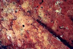 This is a gross photograph of the cut section of the patient's lung showing evidence of severe metastatic calcification. The lung tissue has a rough, firm appearance with open airways.
 -
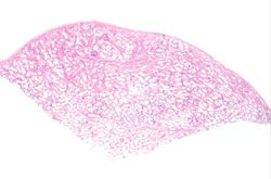 This low-power photomicrograph of the patient's lung illustrates large, open alveolar spaces. The pleural surface is the curved surface at the top.
 -
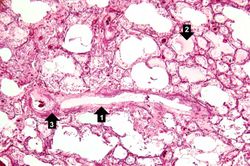 A higher-power photomicrograph shows a blood vessel cut in longitudinal section (1). Several of the alveoli are filled with a pink-staining proteinaceous fluid (2) indicative of pulmonary edema. The alveolar septa and the wall of the blood vessel have a purplish color due to massive deposition of mineral (primarily calcium) in these tissues (3).
 -
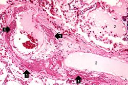 This high-power photomicrograph of a blood vessel shows calcium deposits in the vascular wall (1) and proteinaceous material (2) (from edema) within some of the alveoli. The smooth muscle in the vessel wall has been almost completely replaced by calcium deposits.
 -
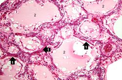 This photomicrograph demonstrates pulmonary alveoli with extensive calcium depositions (1) in the septa and protein accumulations (2) in the alveoli.
 -
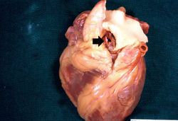 Metastatic calcification is only one of two forms of pathologic calcification. Unlike metastatic calcification, dystrophic calcification does not require an increase in serum calcium levels. This is a gross specimen of a heart with dystrophic calcification of the aortic valve (arrow).
 -
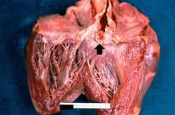 A closer view of this same aortic valve (arrow) illustrates the nodularity and thickening of this valve. This valve would be extremely stiff and almost entirely immobile. This particular example of dystrophic calcification is associated with a degenerative change of the aortic valve due to an unknown cause.
 -
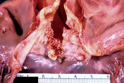 This gross photograph affords a closer view of the same aortic valve. Note the nodularity and thickening of this valve due to fibrosis and dystrophic calcification.


Study Questions
| |||||