Difference between revisions of "IPLab:Lab 11:Leishmaniasis"
Seung Park (talk | contribs) (→Images) |
Seung Park (talk | contribs) (→Related IPLab Cases) |
||
| Line 31: | Line 31: | ||
* [http://peir.path.uab.edu/library/index.php?/tags/2160-leishmaniasis PEIR Digital Library: Leishmaniasis Images] | * [http://peir.path.uab.edu/library/index.php?/tags/2160-leishmaniasis PEIR Digital Library: Leishmaniasis Images] | ||
* [http://library.med.utah.edu/WebPath/INFLHTML/INFLIDX.html WebPath: Inflammation] | * [http://library.med.utah.edu/WebPath/INFLHTML/INFLIDX.html WebPath: Inflammation] | ||
| − | |||
| − | |||
| − | |||
{{IPLab 11}} | {{IPLab 11}} | ||
[[Category: IPLab:Lab 11]] | [[Category: IPLab:Lab 11]] | ||
Revision as of 04:33, 25 August 2013
Contents
Clinical Summary[edit]
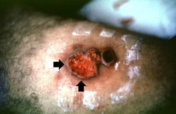
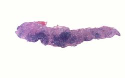
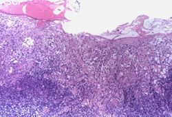
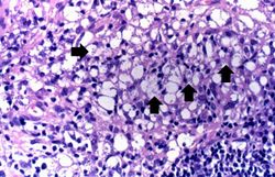
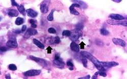
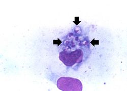
This 28-year-old white male presented to the dermatology clinic complaining of sores on his arms. On examination, two lesions measuring 0.7 to 1.5 cm in diameter were present on his right arm. These lesions showed central ulceration and a raised, indurated margin surrounding the ulcer. The lesions had developed over approximately one month. The patient had applied topical antibiotics, which had no effect. The patient had recently returned from a World Wildlife Fund study site in the Amazon region of Brazil, where he had been conducting field research in the rain forest. A biopsy was taken from the raised edge of one of the ulcers.
Images[edit]
Study Questions[edit]
Additional Resources[edit]
Reference[edit]
- eMedicine Medical Library: Leishmaniasis
- eMedicine Medical Library: Leishmaniasis in Emergency Medicine
- eMedicine Medical Library: Dermatologic Manifestations of Leishmaniasis
- Merck Manual: Leishmaniasis
Journal Articles[edit]
- Choi CM, Lerner EA. Leishmaniasis as an emerging infection. J Investig Dermatol Symp Proc 2001 Dec;6(3):175-82.
Images[edit]
| |||||
An infiltrate is an accumulation of cells in the lung parenchyma--this is a sign of pneumonia.
