Cytologically Yours: CoW: 20140213
Contents
Clinical Summary
The patient is a 66yo old male presenting with jaundice.
Past Medical History
- History of prostate cancer 5 years ago.
- Hyperlipidemia
- Chronic back pain
Past Surgical History
- Prostatectomy (2009)
- Thyroidectomy (2004)
Ultrasound
- Ultrasound of head of pancreas shows a 43mm x 23mm ill defined mass.
Clinical Plan
The differential diagnosis included pancreatic adenocarcinoma and metastatic prostate cancer.
Pathology
Cytology
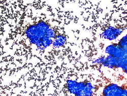
10x magnification of large cohesive groups of cells.

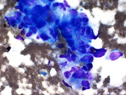
40x magnification showing atypical cells with large irregular nuclei.
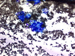
20x magnification showing atypical cells with large irregular nuclei.
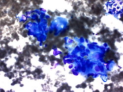
40x magnification showing large, markedly pleomorphic cells.

40x magnification of malignant cell groups.
60x magnification of malignant cells. Macronucleoli are easily identified.


Resident Questions
Cell Block
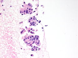
40x magnification of cell block of pancreatic mass showing small glands with macronucleoli.

Final Diagnosis
Cytology
- Metastatic prostate carcinoma.
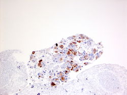
20x magnification of PSA shows positive staining

- CK7 Negative
- CA19.9 Negative
- PSA Positive
Discussion
Metastatic prostate carcinoma to the pancreas is rare; however, long term survival of prostate cancer may increase the incidence. Renal cell carcinoma is the most metastatic pancreatic tumor.