Cytologically Yours: CoW: 20140206
Contents
Clinical Summary
The patient is a 12 year old female with a six month history of left shoulder pain. The patient had tried Aleve and had several chiropractic visits which were unsuccessful at relieving the pain.
Past Medical History
- Previously heathy
Past Surgical History
- No surgical history
Radiology
- AP/Lateral images show a destructive and aggressive appearing lesion in the left proximal huerus in the metaphysis extending 7.5cm distally in the diaphysis.
Clinical Plan
The differential diagnosis included osteosarcoma and Ewing sarcoma. MRI and CT guided biopsy are scheduled.
Pathology
Cytology
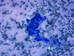
40x magnification of highly atypical malignant appearing cells.
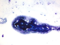
20x magnification showing osteoid formation.
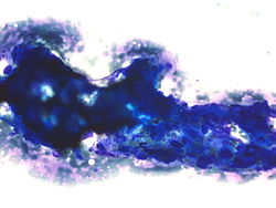
40x magnification showing osteoid formation and malignant appearing cells.
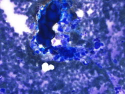
40x magnification of osteoid.


Resident Questions
Final Diagnosis
Cytology
- High grade sarcoma, favor osteosarcoma.
Biopsy
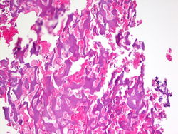
20x magnification of bone biopsy showing sclerotic bone.

Biopsy Diagnosis
- Conventional high grade sarcoma, sclerotic type.
Discussion
The experience of FNA of osteosarcoma is mainly with conventional high-grade intramedullary sarcoma and to the rare high-grade surface osteosarcoma. Smears usually contain dissociated neoplastic cells and cell clusters.
| ||||||||