Cytologically Yours: CoW: 20140120
Contents
Clinical Summary
The patient is a 46 year old female with a new pleural effusion, pelvic mass, and ascites. She presented to the ED with increasing abdominal distension and discomfort. She also has nausea and has been unable to tolerate anything by mouth. She has shortness of breath and has had a recent >20lb weight loss in the last two months.
Past Medical History
- Hyperlipidemia
- Coronary artery disease
- Hepatitis C
Past Surgical History
- Cardiac stent (2011)
- Exploratory pelvic surgery
CT
- Ascites
- Left hydronephrosis
- enlarged uterus 12 x 7 x 7 cm.
- 13 cm pelvic mass
- Right sided pleural effusion
Clinical Plan
Therapeutic paracentesis.
Pathology
Cytology
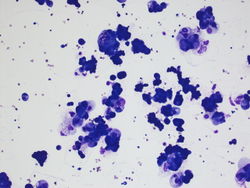
20x magnification of cohesive pleomorphic cells with abundant cytoplasm.(DQ)
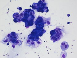
40x magnification showing large atypical cells with abundant cytoplasm. (DQ)
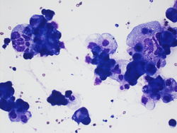
40x magnification showing large groups of cohesive cells that are pleomorphic. (DQ)
40x magnification showing cells that are large and pleomorphic and in groups.(DQ)
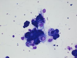
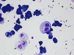
40x magnification of large atypical cells one nucleus appears to have an inclusion.(DQ)
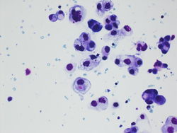
40x magnification of atypical cells. Some of the cells appear to have material in their cytoplasm .(DQ)


Resident Questions
Cell Block
40x magnification cell block
20x magnification cell block


Final Diagnosis
Cytology
- Adenocarcinoma.
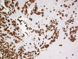
10x magnification of CK7
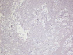
10x magnification of CK20
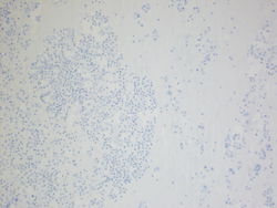
10x magnification of CDX2
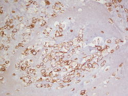
10x magnification of CA125
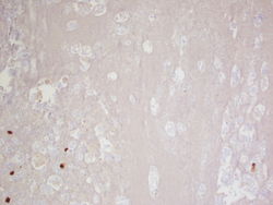
10x magnification of Calretinin


Surgical Pathology
- Metastatic adenocarcinoma, consistent tiwht papillary serous carcinoma.
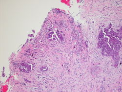
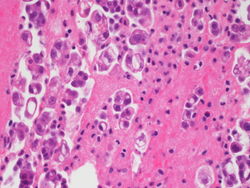
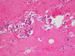
10x magnification of pleural biopsy specimen
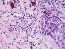
40x magnification of pleural biopsy specimen. Notice the psammoma bodies present.


Discussion
Serous adenocarcinoma commoly presents with widespread peritoneal metastases. Microscopically the tumor can be papillary, solid or nested. Psammoma bodies may be present.