Difference between revisions of "IPLab:Lab 8:Poliomyelitis"
Seung Park (talk | contribs) |
Seung Park (talk | contribs) (→Virtual Microscopy) |
||
| Line 17: | Line 17: | ||
== Virtual Microscopy == | == Virtual Microscopy == | ||
| + | === Poliomyelitis === | ||
<peir-vm>IPLab8Polio</peir-vm> | <peir-vm>IPLab8Polio</peir-vm> | ||
| + | |||
| + | === Normal Spinal Cord === | ||
| + | <peir-vm>IPLab8Polio-Normal_cord</peir-vm> | ||
== Study Questions == | == Study Questions == | ||
Latest revision as of 16:30, 3 January 2014
Contents
Clinical Summary[edit]
Six days before his death, this 31-year-old white male became acutely ill with fever followed by an ascending paralysis which began in his feet. Three days later he was hospitalized because of difficulty in breathing. A lumbar puncture was performed and the patient's spinal fluid contained increased protein and polymorphonuclear leukocytes (4.30 PMNs/mm³). He died on the third hospital day.
Autopsy Findings[edit]
At autopsy, the thoracic and lumbar portions of the spinal cord were softer than normal and focally hemorrhagic.
Images[edit]
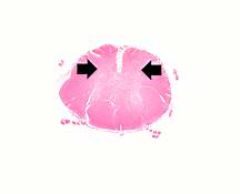
This is a low-power photomicrograph of a section of spinal cord from this case. Note that the anterior horns (arrows) are almost completely devoid of neurons.
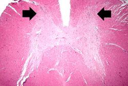
This is a higher-power photomicrograph of spinal cord from this case. Note the absence of motor neurons in the anterior horns and the gliosis (arrows).
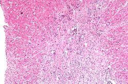
This is a high-power photomicrograph of the anterior horn of the spinal cord from this case. Note the absence of motor neurons and the diffuse gliosis.
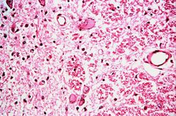
This is a higher-power photomicrograph taken at the junction of the white and gray matter. Note the inflammatory cellular infiltrate and tissue breakdown. There is significant loss of neurons and myelin in this area.
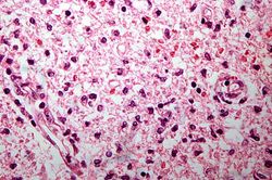
This is another high-power photomicrograph of the anterior horn with inflammatory cell infiltrate and total loss of neurons.


Virtual Microscopy[edit]
Poliomyelitis[edit]
Normal Spinal Cord[edit]
Study Questions[edit]
Additional Resources[edit]
Reference[edit]
- eMedicine Medical Library: Pediatric Poliomyelitis
- Merck Manual: Poliomyelitis
- The WHO: Global Polio Eradication Initiative
Journal Articles[edit]
- Alexander L, Birkhead G, Guerra F, Helms C, Hinman A, Katz S, LeBaron CW, Modlin J, Murphy TV; National Vaccine Advisory Committee-Advisory Committee on Immunization Practices Joint Working Group; Centers for Disease Control and Prevention. Ensuring preparedness for potential poliomyelitis outbreaks: recommendations for the US poliovirus vaccine stockpile from the National Vaccine Advisory Committee (NVAC) and the Advisory Committee on Immunization Practices (ACIP). Arch Pediatr Adolesc Med 2004 Dec;158(12):1106-12.
Images[edit]
- PEIR Digital Library: Polio Images
- WebPath: CNS Pathology: Acquired and Congenital Degenerative Diseases
| |||||
Normally, there should be no PMNs in a patient's spinal fluid.
An infiltrate is an accumulation of cells in the lung parenchyma--this is a sign of pneumonia.