File:IPLab5Hemochromatosis11.jpg
Revision as of 14:57, 20 August 2013 by Peter Anderson (talk | contribs) (This is a histologic section of pancreas from this case stained for iron (Prussian blue). Note the accumulation of iron in the parenchymal cells (1). There is also iron in the pancreatic islets (2).)
No higher resolution available.
IPLab5Hemochromatosis11.jpg (684 × 450 pixels, file size: 81 KB, MIME type: image/jpeg)
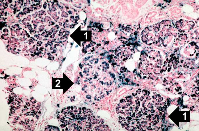
This is a histologic section of pancreas from this case stained for iron (Prussian blue). Note the accumulation of iron in the parenchymal cells (1). There is also iron in the pancreatic islets (2).
File history
Click on a date/time to view the file as it appeared at that time.
| Date/Time | Thumbnail | Dimensions | User | Comment | |
|---|---|---|---|---|---|
| current | 14:57, 20 August 2013 | | 684 × 450 (81 KB) | Peter Anderson (talk | contribs) | This is a histologic section of pancreas from this case stained for iron (Prussian blue). Note the accumulation of iron in the parenchymal cells (1). There is also iron in the pancreatic islets (2). |
- You cannot overwrite this file.
File usage
The following page links to this file: