File:IPLab2Hyperplasia6.jpg
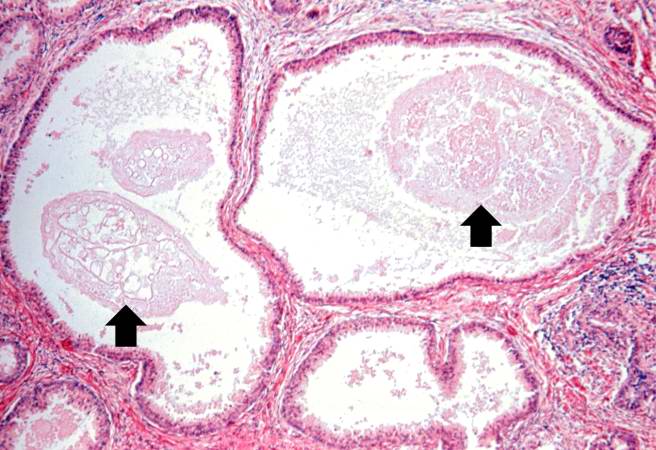
Revision as of 15:28, 19 August 2013 by Peter Anderson (talk | contribs) (Cystic dilatation of glands is present in this photomicrograph. Notice the accumulation of secretory material inside the glands (arrows) and compression (thinning) of the lining epithelium.)
No higher resolution available.
IPLab2Hyperplasia6.jpg (656 × 450 pixels, file size: 61 KB, MIME type: image/jpeg)
Cystic dilatation of glands is present in this photomicrograph. Notice the accumulation of secretory material inside the glands (arrows) and compression (thinning) of the lining epithelium.
File history
Click on a date/time to view the file as it appeared at that time.
| Date/Time | Thumbnail | Dimensions | User | Comment | |
|---|---|---|---|---|---|
| current | 15:28, 19 August 2013 | | 656 × 450 (61 KB) | Peter Anderson (talk | contribs) | Cystic dilatation of glands is present in this photomicrograph. Notice the accumulation of secretory material inside the glands (arrows) and compression (thinning) of the lining epithelium. |
- You cannot overwrite this file.
File usage
The following page links to this file: