File:IPLab2Hyperplasia1.jpg
Revision as of 15:27, 19 August 2013 by Peter Anderson (talk | contribs) (This photograph shows the autopsy specimen from this patient. Included are kidneys (1), ureters (2), bladder (3) (which has been opened), and enlarged prostate (4). Note that the bladder mucosa has multiple trabeculae and the bladder mucosa is hyperemi...)
No higher resolution available.
IPLab2Hyperplasia1.jpg (690 × 450 pixels, file size: 58 KB, MIME type: image/jpeg)
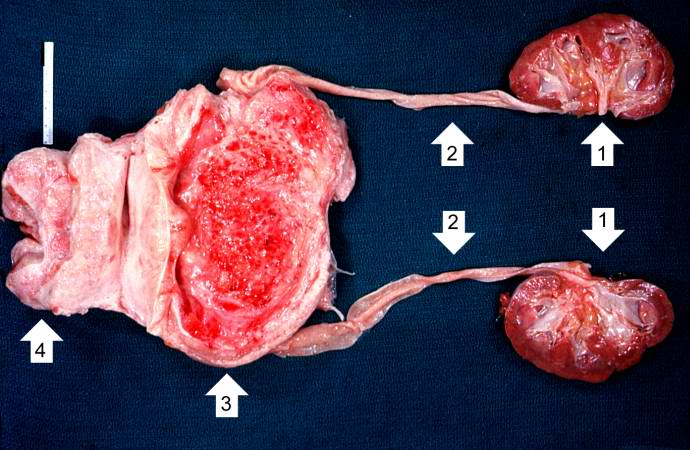
This photograph shows the autopsy specimen from this patient. Included are kidneys (1), ureters (2), bladder (3) (which has been opened), and enlarged prostate (4). Note that the bladder mucosa has multiple trabeculae and the bladder mucosa is hyperemic. Also note that the ureters are dilated.
File history
Click on a date/time to view the file as it appeared at that time.
| Date/Time | Thumbnail | Dimensions | User | Comment | |
|---|---|---|---|---|---|
| current | 15:27, 19 August 2013 | | 690 × 450 (58 KB) | Peter Anderson (talk | contribs) | This photograph shows the autopsy specimen from this patient. Included are kidneys (1), ureters (2), bladder (3) (which has been opened), and enlarged prostate (4). Note that the bladder mucosa has multiple trabeculae and the bladder mucosa is hyperemi... |
- You cannot overwrite this file.
File usage
The following page links to this file: