Difference between revisions of "File:IPLab1MyocardialInfarction4.jpg"
Seung Park (talk | contribs) |
Seung Park (talk | contribs) |
||
| Line 1: | Line 1: | ||
| − | + | This high-power photomicrograph shows the area of infarction on the right (1). There is an area of vacuolated myocytes (2) adjacent to the infarcted myocytes and then normal cardiac muscle to the left (3). | |
Latest revision as of 19:02, 23 August 2013
This high-power photomicrograph shows the area of infarction on the right (1). There is an area of vacuolated myocytes (2) adjacent to the infarcted myocytes and then normal cardiac muscle to the left (3).
File history
Click on a date/time to view the file as it appeared at that time.
| Date/Time | Thumbnail | Dimensions | User | Comment | |
|---|---|---|---|---|---|
| current | 13:50, 15 August 2013 | 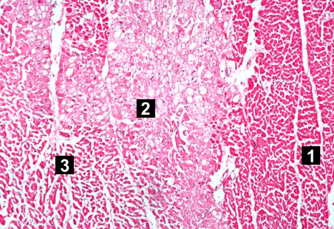 | 655 × 450 (85 KB) | Seung Park (talk | contribs) |
- You cannot overwrite this file.
File usage
The following page links to this file: