Difference between revisions of "File:IPLab1FatNecrosis3.jpg"
Seung Park (talk | contribs) |
Seung Park (talk | contribs) |
||
| Line 1: | Line 1: | ||
| − | + | This low-power photomicrograph of the pancreas from this case shows the fat tissue (1) surrounding the pancreas. Note the rim of inflammatory cells (arrows) and the blue areas in the fat adjacent to the pancreas (2). | |
Latest revision as of 01:40, 16 August 2013
This low-power photomicrograph of the pancreas from this case shows the fat tissue (1) surrounding the pancreas. Note the rim of inflammatory cells (arrows) and the blue areas in the fat adjacent to the pancreas (2).
File history
Click on a date/time to view the file as it appeared at that time.
| Date/Time | Thumbnail | Dimensions | User | Comment | |
|---|---|---|---|---|---|
| current | 01:18, 16 August 2013 | 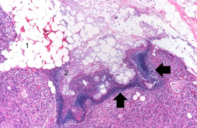 | 691 × 450 (65 KB) | Seung Park (talk | contribs) |
- You cannot overwrite this file.
File usage
The following page links to this file: