Difference between revisions of "File:IPLab12RadiationChanges6.jpg"
Seung Park (talk | contribs) (This is a high-power photomicrograph of the wall of the ileum showing a blood vessel that has suffered radiation-induced damage and is completely occluded (arrows).) |
Seung Park (talk | contribs) |
||
| Line 1: | Line 1: | ||
| − | This | + | This high-power photomicrograph of the wall of the ileum shows areas of fibrosis (1), inflammatory cells (2), and abnormal pleomorphic cells (3) in the area of radiation injury. The abnormal morphology of these cells is radiation-induced. These cells are often difficult to distinguish from recurrent tumor cells. |
Latest revision as of 05:29, 21 August 2013
This high-power photomicrograph of the wall of the ileum shows areas of fibrosis (1), inflammatory cells (2), and abnormal pleomorphic cells (3) in the area of radiation injury. The abnormal morphology of these cells is radiation-induced. These cells are often difficult to distinguish from recurrent tumor cells.
File history
Click on a date/time to view the file as it appeared at that time.
| Date/Time | Thumbnail | Dimensions | User | Comment | |
|---|---|---|---|---|---|
| current | 05:28, 21 August 2013 | 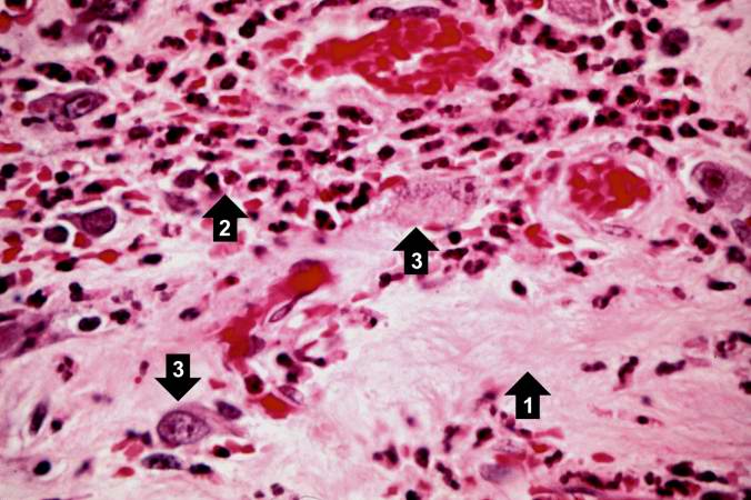 | 676 × 450 (57 KB) | Seung Park (talk | contribs) | This is a high-power photomicrograph of the wall of the ileum showing a blood vessel that has suffered radiation-induced damage and is completely occluded (arrows). |
- You cannot overwrite this file.
File usage
The following page links to this file: