File:IPLab7Carcinoid4.jpg
Revision as of 02:07, 21 August 2013 by Seung Park (talk | contribs) (This is a high-power photomicrograph of the surgical specimen showing the cellular morphology. The tumor cells are monotonously similar with scant, pink, granular cytoplasm and a round-to-oval stippled nucleus. As in most carcinoid tumors, there is min...)
No higher resolution available.
IPLab7Carcinoid4.jpg (667 × 450 pixels, file size: 77 KB, MIME type: image/jpeg)
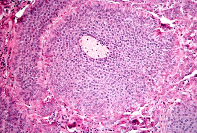
This is a high-power photomicrograph of the surgical specimen showing the cellular morphology. The tumor cells are monotonously similar with scant, pink, granular cytoplasm and a round-to-oval stippled nucleus. As in most carcinoid tumors, there is minimal variation in cell and nuclear size, and mitoses are infrequent or absent.
File history
Click on a date/time to view the file as it appeared at that time.
| Date/Time | Thumbnail | Dimensions | User | Comment | |
|---|---|---|---|---|---|
| current | 02:07, 21 August 2013 | | 667 × 450 (77 KB) | Seung Park (talk | contribs) | This is a high-power photomicrograph of the surgical specimen showing the cellular morphology. The tumor cells are monotonously similar with scant, pink, granular cytoplasm and a round-to-oval stippled nucleus. As in most carcinoid tumors, there is min... |
- You cannot overwrite this file.
File usage
The following page links to this file: