File:IPLab2Calcification2.jpg
Revision as of 16:38, 19 August 2013 by Peter Anderson (talk | contribs) (This low-power photomicrograph of the patient's lung illustrates large, open alveolar spaces. The pleural surface is the curved surface at the top.)
No higher resolution available.
IPLab2Calcification2.jpg (683 × 450 pixels, file size: 37 KB, MIME type: image/jpeg)
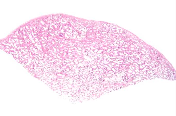
This low-power photomicrograph of the patient's lung illustrates large, open alveolar spaces. The pleural surface is the curved surface at the top.
File history
Click on a date/time to view the file as it appeared at that time.
| Date/Time | Thumbnail | Dimensions | User | Comment | |
|---|---|---|---|---|---|
| current | 16:38, 19 August 2013 | | 683 × 450 (37 KB) | Peter Anderson (talk | contribs) | This low-power photomicrograph of the patient's lung illustrates large, open alveolar spaces. The pleural surface is the curved surface at the top. |
- You cannot overwrite this file.
File usage
The following page links to this file: