File:IPLab2Hyperplasia2.jpg
Revision as of 15:27, 19 August 2013 by Peter Anderson (talk | contribs) (This is a close-up of the prostate from this same patient. Note the nodularity of the tissue (1) and the enlargement of the gland. Enlargement of the prostate leads to compression of the urethra as it passes through (2) the gland.)
No higher resolution available.
IPLab2Hyperplasia2.jpg (654 × 450 pixels, file size: 44 KB, MIME type: image/jpeg)
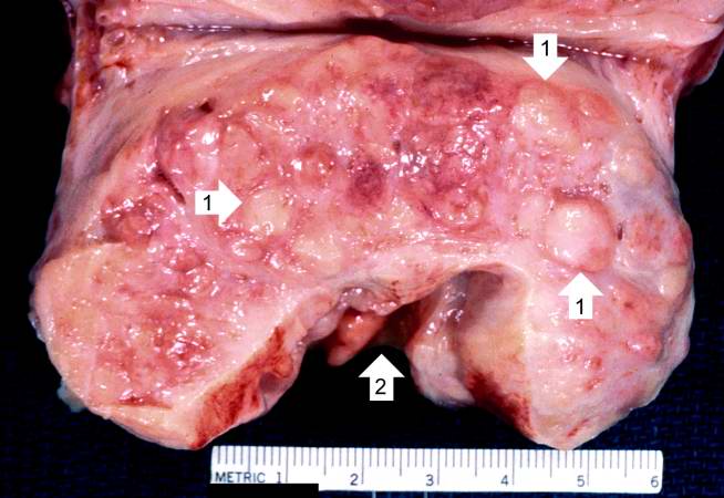
This is a close-up of the prostate from this same patient. Note the nodularity of the tissue (1) and the enlargement of the gland. Enlargement of the prostate leads to compression of the urethra as it passes through (2) the gland.
File history
Click on a date/time to view the file as it appeared at that time.
| Date/Time | Thumbnail | Dimensions | User | Comment | |
|---|---|---|---|---|---|
| current | 15:27, 19 August 2013 | | 654 × 450 (44 KB) | Peter Anderson (talk | contribs) | This is a close-up of the prostate from this same patient. Note the nodularity of the tissue (1) and the enlargement of the gland. Enlargement of the prostate leads to compression of the urethra as it passes through (2) the gland. |
- You cannot overwrite this file.
File usage
The following page links to this file: