File:IPLab12RadiationChanges4.jpg
Revision as of 05:28, 21 August 2013 by Seung Park (talk | contribs) (This is a high-power photomicrograph showing the atrophied epithelium in the area of radiation injury (1). Note the dense fibrous connective tissue within the wall of the ileum and the congested blood vessels (2).)
No higher resolution available.
IPLab12RadiationChanges4.jpg (298 × 450 pixels, file size: 39 KB, MIME type: image/jpeg)
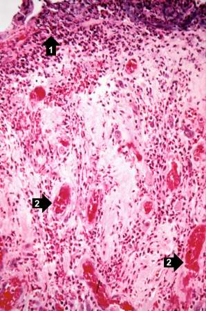
This is a high-power photomicrograph showing the atrophied epithelium in the area of radiation injury (1). Note the dense fibrous connective tissue within the wall of the ileum and the congested blood vessels (2).
File history
Click on a date/time to view the file as it appeared at that time.
| Date/Time | Thumbnail | Dimensions | User | Comment | |
|---|---|---|---|---|---|
| current | 05:28, 21 August 2013 | | 298 × 450 (39 KB) | Seung Park (talk | contribs) | This is a high-power photomicrograph showing the atrophied epithelium in the area of radiation injury (1). Note the dense fibrous connective tissue within the wall of the ileum and the congested blood vessels (2). |
- You cannot overwrite this file.
File usage
The following page links to this file: