File:IPLab12Alcoholic4.jpg
Revision as of 05:13, 21 August 2013 by Seung Park (talk | contribs) (In this is medium-power photomicrograph of trichrome stained liver the bands of fibrous tissue are seen to form "bridges" between triad areas (arrows); this is called "bridging fibrosis." Also note the fibrous tissue (arrows) and how the hepatocytes ar...)
No higher resolution available.
IPLab12Alcoholic4.jpg (685 × 450 pixels, file size: 130 KB, MIME type: image/jpeg)
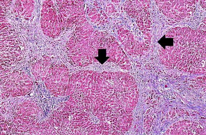
In this is medium-power photomicrograph of trichrome stained liver the bands of fibrous tissue are seen to form "bridges" between triad areas (arrows); this is called "bridging fibrosis." Also note the fibrous tissue (arrows) and how the hepatocytes are separated into nodules by this fibrous tissue.
File history
Click on a date/time to view the file as it appeared at that time.
| Date/Time | Thumbnail | Dimensions | User | Comment | |
|---|---|---|---|---|---|
| current | 05:13, 21 August 2013 | | 685 × 450 (130 KB) | Seung Park (talk | contribs) | In this is medium-power photomicrograph of trichrome stained liver the bands of fibrous tissue are seen to form "bridges" between triad areas (arrows); this is called "bridging fibrosis." Also note the fibrous tissue (arrows) and how the hepatocytes ar... |
- You cannot overwrite this file.
File usage
The following page links to this file: