File:IPLab11Malaria7.jpg
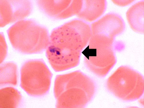
Revision as of 04:56, 21 August 2013 by Seung Park (talk | contribs) (In this peripheral smear from a different patient who was infected with P. vivax, the cytoplasm of the infected RBC has a stippled appearance (Schüffner's dots) (arrow). The RBC is also slightly enlarged.)
No higher resolution available.
IPLab11Malaria7.jpg (601 × 450 pixels, file size: 24 KB, MIME type: image/jpeg)
In this peripheral smear from a different patient who was infected with P. vivax, the cytoplasm of the infected RBC has a stippled appearance (Schüffner's dots) (arrow). The RBC is also slightly enlarged.
File history
Click on a date/time to view the file as it appeared at that time.
| Date/Time | Thumbnail | Dimensions | User | Comment | |
|---|---|---|---|---|---|
| current | 04:56, 21 August 2013 | | 601 × 450 (24 KB) | Seung Park (talk | contribs) | In this peripheral smear from a different patient who was infected with P. vivax, the cytoplasm of the infected RBC has a stippled appearance (Schüffner's dots) (arrow). The RBC is also slightly enlarged. |
- You cannot overwrite this file.
File usage
The following page links to this file: