File:IPLab7Melanoma8.jpg
Revision as of 01:57, 21 August 2013 by Seung Park (talk | contribs) (This is a high-power photomicrograph of the main tumor mass showing the cellular details. The individual melanoma cells contain large nuclei with irregular contours having chromatin clumped at the periphery of the nuclear membrane and prominent red (eo...)
No higher resolution available.
IPLab7Melanoma8.jpg (690 × 450 pixels, file size: 76 KB, MIME type: image/jpeg)
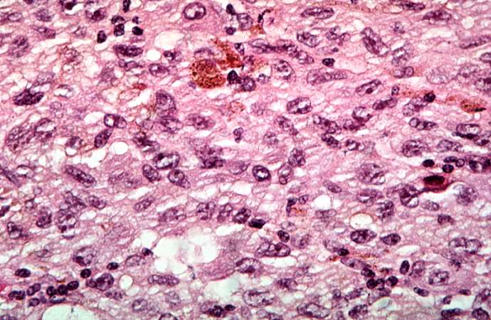
This is a high-power photomicrograph of the main tumor mass showing the cellular details. The individual melanoma cells contain large nuclei with irregular contours having chromatin clumped at the periphery of the nuclear membrane and prominent red (eosinophilic) nucleoli.
File history
Click on a date/time to view the file as it appeared at that time.
| Date/Time | Thumbnail | Dimensions | User | Comment | |
|---|---|---|---|---|---|
| current | 01:57, 21 August 2013 | | 690 × 450 (76 KB) | Seung Park (talk | contribs) | This is a high-power photomicrograph of the main tumor mass showing the cellular details. The individual melanoma cells contain large nuclei with irregular contours having chromatin clumped at the periphery of the nuclear membrane and prominent red (eo... |
- You cannot overwrite this file.
File usage
The following page links to this file: