File:IPLab7ColonCA1.jpg
Revision as of 01:40, 21 August 2013 by Seung Park (talk | contribs) (This is a gross photograph of the adenoma from the surgical specimen in this case. Note the large, ulcerated, fungating annular (encircling) carcinoma (1) with areas of hemorrhage (2). Also note the adenomatous polyps (3).)
No higher resolution available.
IPLab7ColonCA1.jpg (673 × 450 pixels, file size: 69 KB, MIME type: image/jpeg)
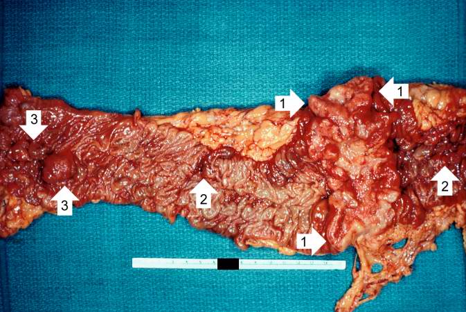
This is a gross photograph of the adenoma from the surgical specimen in this case. Note the large, ulcerated, fungating annular (encircling) carcinoma (1) with areas of hemorrhage (2). Also note the adenomatous polyps (3).
File history
Click on a date/time to view the file as it appeared at that time.
| Date/Time | Thumbnail | Dimensions | User | Comment | |
|---|---|---|---|---|---|
| current | 01:40, 21 August 2013 | | 673 × 450 (69 KB) | Seung Park (talk | contribs) | This is a gross photograph of the adenoma from the surgical specimen in this case. Note the large, ulcerated, fungating annular (encircling) carcinoma (1) with areas of hemorrhage (2). Also note the adenomatous polyps (3). |
- You cannot overwrite this file.
File usage
The following page links to this file: