IPLab:Lab 3:Tuberculosis
Clinical Summary
Two months prior to admission, this 57-year-old white male underwent a routine physical examination. During the course of the examination, a lesion was found in the upper lobe of the right lung. Initially, the patient was treated for two weeks with ampicillin. He was then admitted to the hospital for further study. All studies including sputum studies for tubercle bacilli, bronchial washings, and bronchoscopy were negative and he was discharged.
Upon re-admission, a review of systems revealed the presence of mild dyspnea on exertion, accompanied by a slightly productive cough. Of interest was the fact that the patient had had a positive PPD test for the past 4 to 5 years, but this had never been evaluated. At this time, physical and laboratory examinations were unremarkable. Radiographic examination of the chest revealed a 2 x 2-cm density in the right lower lung field. A CT scan revealed several small cavities in this area.
Images
-
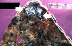 This is a gross photograph of a lung containing a nodular lesion at the lung apex (arrows). Note that the lesion appears solid and has a whitish coloration indicating considerable fibrous connective tissue. This is a healed granuloma due to primary tuberculosis in the lung. There are smaller focal lesions adjacent to the major mass. In addition, note the extensive anthracosis in this lung.
 -
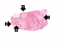 This low-power photomicrograph of a section of lung reveals multiple large nodules (1) with pale eosinophilic centers surrounded by a rim of blue-staining nuclei. In addition to the large nodules, there are several smaller nodules throughout the slide (2).
 -
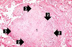 This is a photomicrograph of a tuberculosis granuloma. Note the central core of caseation necrosis (1) encircled by a rim of epithelioid macrophages and lymphocytes (2). Langhans’ type multinucleated giant cells are also present although they are difficult to see at this power (3).
 -
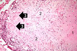 This is a high-power photomicrograph of a granuloma. Note the necrotic core on the right (1), epithelioid macrophages (2), and Langhans’ type giant cells (3) at the periphery of the granuloma. Note also the small lymphocytes characterized by their distinctly blue-staining nuclei. Other cells in the tissue, in addition to the macrophages, include fibroblasts and occasional neutrophils.
 -
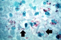 This high-power photomicrograph of a tuberculosis granuloma demonstrates acid-fast bacilli (arrows).


Virtual Microscopy
Lung: Tuberculosis H&E
Lung: Tuberculosis AFGT
Normal Lung
Study Questions
Additional Resources
Reference
Journal Articles
- Rodrigues DS, Medeiros EA, Weckx LY, Bonnez W, Salomão R, Kallas EG. Immunophenotypic characterization of peripheral T lymphocytes in Mycobacterium tuberculosis infection and disease. Clin Exp Immunol 2002 Apr;128(1):149-54.
- Horsburgh CR, Barry CE, Lange C. Treatment of Tuberculosis. NEJM 2015 Nov 26;373:2149-2160.