IPLab:Lab 4:Pulmonary Congestion and Edema
Clinical Summary
This 67-year-old male was hospitalized because of extensive atherosclerotic cardiovascular disease. Following surgery, during which diseased portions of the femoral arteries were bypassed, he developed massive pulmonary embolization and expired.
At autopsy, thrombi were found in the femoral and iliac veins, as well as in the larger pulmonary arteries.
Autopsy Findings
Significant findings at postmortem examination were old and recent myocardial infarctions and evidence of congestive heart failure. The right and left lungs weighed 950 grams and 750 grams, respectively, and were reddish-brown.
Images
-
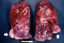 This is a gross photograph of lungs that are distended and red. The reddish coloration of the tissue is due to congestion. Some normal pink lung tissue is seen at the edges of the lungs (arrows).
 -
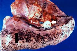 This is a gross photograph of lung demonstrating acute pulmonary congestion and edema. A frothy exudate fills the bronchus (arrow).
 -
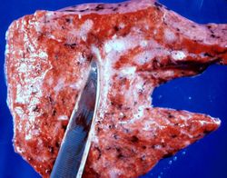 This gross photograph demonstrates the frothy exudate that is being extruded from the lung tissue.
 -
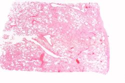 This is a low-power photomicrograph of lung from this case. The lung section has a pale-red color indicating proteinaceous material within the lung.
 -
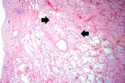 This is a higher-power photomicrograph of lung. The edema fluid within the alveoli is visible at this higher magnification (arrows). The thickened pleura (1) is on the left.
 -
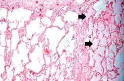 This is a higher-power photomicrograph showing edema-filled alveoli in the right portion of this section (arrows).
 -
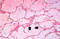 This high-power photomicrograph illustrates the edema fluid within the alveoli (1) and the congestion (RBCs) in the alveolar capillaries (arrows).


Virtual Microscopy
Lung: Congestion and Edema
Normal Lung
Study Questions
- Define the following:
Additional Resources
Reference
Journal Articles
- Welch TD, Yang EH, Reeder GS, Gersh BJ. Modern management of acute myocardial infarction. Curr Probl Cardiol 2012 Jul;37(7):237-310.
Images
Related IPLab Cases
- Lab 1: Heart: Myocardial Infarction (Coagulative Necrosis)
- Lab 3: Heart: Acute Myocardial Infarction
- Lab 3: Heart: Healed Myocardial Infarction
- Lab 4: Heart: Mural Thrombus
- Lab 4: Coronary Artery: Thrombosis
| |||||