From Pathology Education Instructional Resource
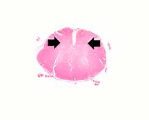
This is a low-power photomicrograph of a section of spinal cord from this case. Note that the anterior horns (arrows) are almost completely devoid of neurons.
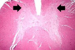
This is a higher-power photomicrograph of spinal cord from this case. Note the absence of motor neurons in the anterior horns and the gliosis (arrows).
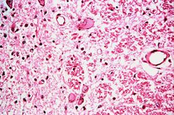
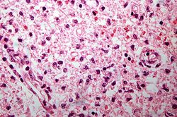
This is a high-power photomicrograph of the anterior horn of the spinal cord from this case. Note the absence of motor neurons and the diffuse gliosis.
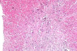
This is a higher-power photomicrograph taken at the junction of the white and gray matter. Note the inflammatory cellular infiltrate and tissue breakdown. There is significant loss of neurons and myelin in this area.
This is another high-power photomicrograph of the anterior horn with inflammatory cell infiltrate and total loss of neurons.
