File:IPLab12Alcoholic6.jpg
Revision as of 05:13, 21 August 2013 by Seung Park (talk | contribs) (In this high-power photomicrograph of trichrome-stained liver, the bands of fibrous tissue surround the hepatocyte nodules. There is some degeneration and dropout of hepatocytes in this nodule. Also note the increased numbers of bile ducts in the triad...)
No higher resolution available.
IPLab12Alcoholic6.jpg (607 × 450 pixels, file size: 42 KB, MIME type: image/jpeg)
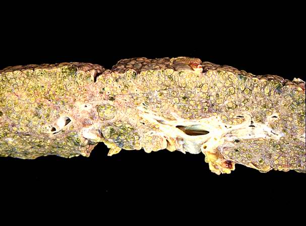
In this high-power photomicrograph of trichrome-stained liver, the bands of fibrous tissue surround the hepatocyte nodules. There is some degeneration and dropout of hepatocytes in this nodule. Also note the increased numbers of bile ducts in the triad area (arrows). Bile duct proliferation is a common feature in many hepatitides.
File history
Click on a date/time to view the file as it appeared at that time.
| Date/Time | Thumbnail | Dimensions | User | Comment | |
|---|---|---|---|---|---|
| current | 05:13, 21 August 2013 | | 607 × 450 (42 KB) | Seung Park (talk | contribs) | In this high-power photomicrograph of trichrome-stained liver, the bands of fibrous tissue surround the hepatocyte nodules. There is some degeneration and dropout of hepatocytes in this nodule. Also note the increased numbers of bile ducts in the triad... |
- You cannot overwrite this file.
File usage
The following page links to this file: