Difference between revisions of "File:IPLab1MyocardialInfarction2.jpg"
Seung Park (talk | contribs) |
Seung Park (talk | contribs) |
||
| Line 1: | Line 1: | ||
| − | + | This is a low-power photomicrograph of the left ventricular free wall extending from the epicardium (1) to the endocardium (2). The area of infarction is the darker red (hypereosinophilic area) along the subendocardium (3). | |
Latest revision as of 14:01, 15 August 2013
This is a low-power photomicrograph of the left ventricular free wall extending from the epicardium (1) to the endocardium (2). The area of infarction is the darker red (hypereosinophilic area) along the subendocardium (3).
File history
Click on a date/time to view the file as it appeared at that time.
| Date/Time | Thumbnail | Dimensions | User | Comment | |
|---|---|---|---|---|---|
| current | 13:49, 15 August 2013 | 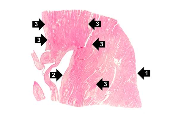 | 609 × 450 (22 KB) | Seung Park (talk | contribs) |
- You cannot overwrite this file.
File usage
The following page links to this file: