IPLab:Lab 4:Chronic Passive Congestion: Difference between revisions
Seung Park (talk | contribs) No edit summary |
Seung Park (talk | contribs) |
||
| Line 35: | Line 35: | ||
=== Images === | === Images === | ||
* [ | * [{{SERVER}}/library/index.php?/tags/672-chronic_passive_congestion PEIR Digital Library: Chronic Passive Congestion Images] | ||
* [http://library.med.utah.edu/WebPath/LIVEHTML/LIVERIDX.html#1 Webpath: Hepatic Pathology] | * [http://library.med.utah.edu/WebPath/LIVEHTML/LIVERIDX.html#1 Webpath: Hepatic Pathology] | ||
Revision as of 20:30, 29 August 2013
Clinical Summary
This 57-year-old male was hospitalized with a three-month history of a dry, hacking cough, dyspnea, and chest pain. He was diagnosed as having congestive heart failure due to viral myocarditis. There was no evidence of myocardial infarction, but renal and hepatic functions were decreased. The patient developed refractory congestive heart failure and renal failure. The patient's cardiac index was marginal and he was classified as a having congestive cardiomyopathy. The patient had a cardiorespiratory arrest and died two months after admission.
Autopsy Findings
At autopsy the lungs were congested and edematous with evidence of lobar pneumonia. The heart weighed 540 grams and showed 75 to 95% atherosclerotic stenosis of all the major coronary arteries. The right and left ventricles were markedly dilated. The ventricular walls were extremely thin with patchy areas of subendocardial fibrosis. The liver weighed 1630 grams and displayed the classic "nutmeg appearance" of chronic passive hepatic congestion on cut surface.
Images
-
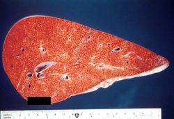 This is a gross photograph of a liver demonstrating chronic passive congestion. Note the accentuation of the centrilobular pattern evidenced by the dark-brown-staining areas in this tissue.
 -
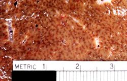 This is a closer view of a cut section of liver demonstrating the pattern of chronic passive congestion. The central vein regions are red and the surrounding hepatic tissue is pale tan-brown.
 -
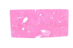 This low-power photomicrograph of liver demonstrates a slightly visible pattern of centrilobular congestion at this magnification.
 -
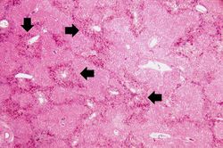 This is a higher-power photomicrograph of liver demonstrating an accentuated lobular pattern with a dark red stain surrounding the central veins in the liver lobules (arrows).
 -
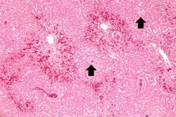 This higher-power photomicrograph of the liver lobules shows congestion and red blood cell accumulation in the sinusoidal spaces around the central vein. Note that around the portal triads (arrows) the liver cells are quite normal and there is no evidence of congestion.
 -
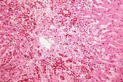 This is a high-power photomicrograph of the central vein illustrating congestion and some loss of liver parenchymal cells. A mild increase in connective tissue around the central vein is evident in this section.
 -
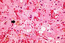 This is a high-power photomicrograph of liver with several macrophages that are distended with a brown pigment (arrow). These resident macrophages (Kupffer cells) are part of the reticuloendothelial system and normally line the sinusoidal spaces in the liver where they phagocytose the RBCs that pool, and eventually die, in the central vein region.
 -
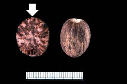 This is a gross photograph of a nutmeg. You can see from the appearance of the cut surface of the nutmeg (arrow) why chronic passive congestion of the liver is sometimes referred to as "nutmeg liver."
 -
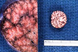 This is a gross photograph of the cut surface of a liver with chronic passive congestion (left) compared to the cut surface of a nutmeg (right).


Study Questions
Additional Resources
Reference
- eMedicine Medical Library: Cardiac Cirrhosis and Congestive Hepatopathy
- eMedicine Medical Library: Myocarditis
- eMedicine Medical Library: Pediatric Congestive Heart Failure
- Merck Manual: Cirrhosis
Journal Articles
- Naschitz JE, Slobodin G, Lewis RJ, Zuckerman E, Yeshurun D. Heart diseases affecting the liver and liver diseases affecting the heart. Am Heart J 2000 Jul;140(1):111-20.
Images
Related IPLab Cases
| |||||