Difference between revisions of "File:IPLab1MyocardialInfarction5.jpg"
Seung Park (talk | contribs) |
Seung Park (talk | contribs) |
||
| Line 1: | Line 1: | ||
| − | + | This high-power photomicrograph shows the endocardium (1) and the area of subendocardial vacuolar degeneration (2). The area of infarction (3) contains some red blood cells. | |
Latest revision as of 19:02, 23 August 2013
This high-power photomicrograph shows the endocardium (1) and the area of subendocardial vacuolar degeneration (2). The area of infarction (3) contains some red blood cells.
File history
Click on a date/time to view the file as it appeared at that time.
| Date/Time | Thumbnail | Dimensions | User | Comment | |
|---|---|---|---|---|---|
| current | 13:50, 15 August 2013 | 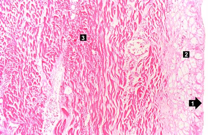 | 694 × 450 (77 KB) | Seung Park (talk | contribs) |
- You cannot overwrite this file.
File usage
The following page links to this file: