IPLab:Lab 8:Rabies: Difference between revisions
Jump to navigation
Jump to search
Seung Park (talk | contribs) |
Seung Park (talk | contribs) No edit summary |
||
| (One intermediate revision by the same user not shown) | |||
| Line 11: | Line 11: | ||
File:IPLab8Rabies5.jpg|This is a high-power photomicrograph of a neuron surrounded by inflammatory cells (lymphocytes and microglia). This neuron has two intracytoplasmic eosinophilic inclusion bodies (arrows). | File:IPLab8Rabies5.jpg|This is a high-power photomicrograph of a neuron surrounded by inflammatory cells (lymphocytes and microglia). This neuron has two intracytoplasmic eosinophilic inclusion bodies (arrows). | ||
</gallery> | </gallery> | ||
== Virtual Microscopy == | |||
<peir-vm>IPLab8Rabies</peir-vm> | |||
== Study Questions == | == Study Questions == | ||
| Line 28: | Line 31: | ||
=== Images === | === Images === | ||
* [ | * [{{SERVER}}/library/index.php?/tags/1652-rabies PEIR Digital Library: Rabies Images] | ||
* [http://library.med.utah.edu/WebPath/CNSHTML/CNSIDX.html#5 WebPath: CNS Infections] | * [http://library.med.utah.edu/WebPath/CNSHTML/CNSIDX.html#5 WebPath: CNS Infections] | ||
Latest revision as of 11:29, 3 January 2014
Clinical Summary
This 52-year-old female had been bitten by a dog two months previously. One week prior to death, she developed severe headache, restlessness, and dysphagia. These symptoms were followed by the appearance of fever, tremor, and general rigidity. The patient was admitted, but expired on the second hospital day.
Images
-
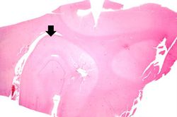 This is a low-power photomicrograph of the hippocampus (arrow) from this case.
 -
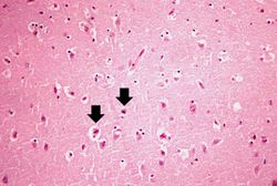 This is a medium-power photomicrograph of brain tissue exhibiting edema and evidence of shrunken, necrotic neurons (arrows).
 -
 This is a higher-power photomicrograph of the shrunken neurons. One neuron appears to have an eosinophilic intracytoplasmic inclusion body (arrow).
 -
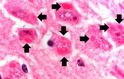 This is a high-power photomicrograph of neurons containing variably-sized intracytoplasmic eosinophilic inclusion bodies (arrows).
 -
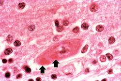 This is a high-power photomicrograph of a neuron surrounded by inflammatory cells (lymphocytes and microglia). This neuron has two intracytoplasmic eosinophilic inclusion bodies (arrows).


Virtual Microscopy
Study Questions
Additional Resources
Reference
- eMedicine Medical Library: Emergency Treatment of Rabies
- eMedicine Medical Library: Rabies
- Merck Manual: Rabies
- Merck Manual: Rabies Postexposure Prophylaxis
Journal Articles
- Jogai S, Radotra BD, Banerjee AK. Immunohistochemical study of human rabies. Neuropathology 2000 Sep;20(3):197-203.
Images
| |||||