From Pathology Education Instructional Resource
Revision as of 02:50, 21 August 2013
Images
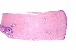
This is a low-power photomicrograph of liver from this case. This section was stained with a modified aldehyde fuchsin and counterstained with hematoxylin and eosin. Modified aldehyde fuchsin colors cystine-rich proteins--such as HBsAg and elastic fibers--deep purple. The cytoplasm of most liver cells (and RBCs) stain red due to the eosin and have dark blue nuclei.
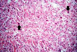
This is a higher-power photomicrograph of liver from this case. Note the severe congestion (RBCs in sinusoids) and the presence of occasional hepatocytes with dark red/magenta-stained cytoplasm (arrows).
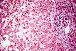
This is a higher-power photomicrograph of the periportal region exhibiting some inflammation and bile duct hyperplasia. There is also congestion and some loss of hepatocytes with disruption of the hepatic cords.
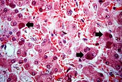
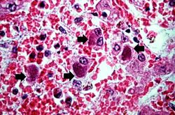
This is a high-power photomicrograph of liver with numerous hepatocytes containing accumulations of magenta-staining material in the cytoplasm (arrows).
This high-power photomicrograph showing hepatocytes, the cytoplasm of which contain intracytoplasmic accumulations (arrows) of hepatitis B surface antigen.
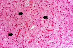
This is a photomicrograph of a liver section from another case of hepatitis B. In this H&E-stained section, the typical "ground glass" appearance of the hepatocytes can be appreciated (arrows).
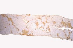
This is a low-power photomicrograph of liver from the previous image which has been reacted with antibody specific for HBsAg. The hepatocytes that contain HBsAg stain brown.
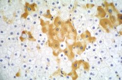
This higher-power photomicrograph of the previous section shows more clearly the HBsAg positive cells (arrows). Upon staining with H&E, these same cells exhibit a "ground glass" appearance, which is due to the accumulation of HBsAg in the hepatocyte cytoplasm.
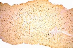
This is a low-power photomicrograph of the same liver reacted with antibody specific for hepatitis B core antigen (HBcAg). The hepatocytes that contain HBcAg stain brown. Note that even at this low magnification, many brown-staining nuclei can be seen.
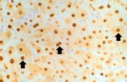
This high-power photomicrograph of the previous section shows the HBcAg positive nuclei (arrows).
