Difference between revisions of "File:IPLab6Hashimoto3.jpg"
(This is a higher-power photomicrograph of thyroid from this case. Note the large number of blue-staining inflammatory cells in this tissue. These cells appear to be forming germinal centers. Some residual thyroid gland tissue can be seen in this sectio...) |
(No difference)
|
Latest revision as of 17:43, 20 August 2013
This is a higher-power photomicrograph of thyroid from this case. Note the large number of blue-staining inflammatory cells in this tissue. These cells appear to be forming germinal centers. Some residual thyroid gland tissue can be seen in this section (arrows).
File history
Click on a date/time to view the file as it appeared at that time.
| Date/Time | Thumbnail | Dimensions | User | Comment | |
|---|---|---|---|---|---|
| current | 17:43, 20 August 2013 | 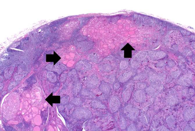 | 671 × 450 (55 KB) | Peter Anderson (talk | contribs) | This is a higher-power photomicrograph of thyroid from this case. Note the large number of blue-staining inflammatory cells in this tissue. These cells appear to be forming germinal centers. Some residual thyroid gland tissue can be seen in this sectio... |
- You cannot overwrite this file.
File usage
The following page links to this file: