Difference between revisions of "File:IPLab5Hemochromatosis6.jpg"
(This trichrome stain of liver section demonstrates the increased fibrous connective tissue in this liver. Note that the liver nodules (1) are surrounded by fibrous connective tissue (2).) |
(No difference)
|
Latest revision as of 14:54, 20 August 2013
This trichrome stain of liver section demonstrates the increased fibrous connective tissue in this liver. Note that the liver nodules (1) are surrounded by fibrous connective tissue (2).
File history
Click on a date/time to view the file as it appeared at that time.
| Date/Time | Thumbnail | Dimensions | User | Comment | |
|---|---|---|---|---|---|
| current | 14:54, 20 August 2013 | 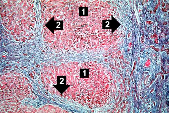 | 673 × 450 (99 KB) | Peter Anderson (talk | contribs) | This trichrome stain of liver section demonstrates the increased fibrous connective tissue in this liver. Note that the liver nodules (1) are surrounded by fibrous connective tissue (2). |
- You cannot overwrite this file.
File usage
The following page links to this file: