Difference between revisions of "File:IPLab2Calcification5.jpg"
(This photomicrograph demonstrates pulmonary alveoli with extensive calcium depositions (1) in the septa and protein accumulations (2) in the alveoli.) |
(No difference)
|
Latest revision as of 16:34, 19 August 2013
This photomicrograph demonstrates pulmonary alveoli with extensive calcium depositions (1) in the septa and protein accumulations (2) in the alveoli.
File history
Click on a date/time to view the file as it appeared at that time.
| Date/Time | Thumbnail | Dimensions | User | Comment | |
|---|---|---|---|---|---|
| current | 16:34, 19 August 2013 | 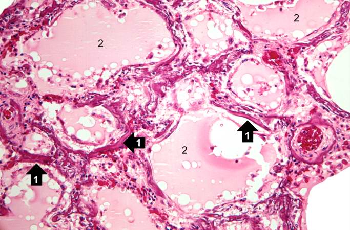 | 683 × 450 (68 KB) | Peter Anderson (talk | contribs) | This photomicrograph demonstrates pulmonary alveoli with extensive calcium depositions (1) in the septa and protein accumulations (2) in the alveoli. |
- You cannot overwrite this file.
File usage
The following page links to this file: