Difference between revisions of "File:IPLab2Hyperplasia3.jpg"
(This is a low-power photomicrograph showing hyperplastic prostate on the left (1) and normal prostate on the right (2). At this power, dilated glands are visible in the section of hyperplastic prostate.) |
(No difference)
|
Latest revision as of 15:28, 19 August 2013
This is a low-power photomicrograph showing hyperplastic prostate on the left (1) and normal prostate on the right (2). At this power, dilated glands are visible in the section of hyperplastic prostate.
File history
Click on a date/time to view the file as it appeared at that time.
| Date/Time | Thumbnail | Dimensions | User | Comment | |
|---|---|---|---|---|---|
| current | 15:28, 19 August 2013 | 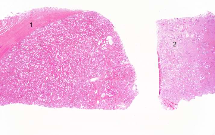 | 714 × 450 (48 KB) | Peter Anderson (talk | contribs) | This is a low-power photomicrograph showing hyperplastic prostate on the left (1) and normal prostate on the right (2). At this power, dilated glands are visible in the section of hyperplastic prostate. |
- You cannot overwrite this file.
File usage
The following page links to this file: