File:IPLab1FatNecrosis6.jpg: Difference between revisions
Jump to navigation
Jump to search
Seung Park (talk | contribs) No edit summary |
Seung Park (talk | contribs) No edit summary |
||
| Line 1: | Line 1: | ||
A higher-power photomicrograph of the previous slide contains a small area of fat necrosis (1) in the upper right portion of the image. The fat necrosis is within the fat tissue that is normally found adjacent to the pancreas. The appearance of the pancreatic tissue in this area is somewhat disrupted due to autolysis (the pancreas autolyzes very rapidly after death) but there is some premortem necrosis as well. | |||


Latest revision as of 20:42, 15 August 2013
A higher-power photomicrograph of the previous slide contains a small area of fat necrosis (1) in the upper right portion of the image. The fat necrosis is within the fat tissue that is normally found adjacent to the pancreas. The appearance of the pancreatic tissue in this area is somewhat disrupted due to autolysis (the pancreas autolyzes very rapidly after death) but there is some premortem necrosis as well.
File history
Click on a date/time to view the file as it appeared at that time.
| Date/Time | Thumbnail | Dimensions | User | Comment | |
|---|---|---|---|---|---|
| current | 20:18, 15 August 2013 | 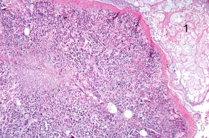 | 677 × 450 (83 KB) | Seung Park (talk | contribs) |
You cannot overwrite this file.
File usage
The following page uses this file:
