Difference between revisions of "File:IPLab13WT6.jpg"
Seung Park (talk | contribs) (This medium-power photomicrograph of tumor shows again the two cell types making up this neoplasm. There are regions within the blastema where the cells form glands or "tubules" (arrows).) |
(No difference)
|
Latest revision as of 05:55, 21 August 2013
This medium-power photomicrograph of tumor shows again the two cell types making up this neoplasm. There are regions within the blastema where the cells form glands or "tubules" (arrows).
File history
Click on a date/time to view the file as it appeared at that time.
| Date/Time | Thumbnail | Dimensions | User | Comment | |
|---|---|---|---|---|---|
| current | 05:55, 21 August 2013 | 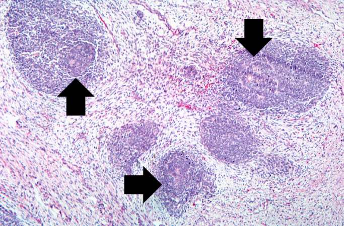 | 683 × 450 (84 KB) | Seung Park (talk | contribs) | This medium-power photomicrograph of tumor shows again the two cell types making up this neoplasm. There are regions within the blastema where the cells form glands or "tubules" (arrows). |
- You cannot overwrite this file.
File usage
The following page links to this file: