File list
This special page shows all uploaded files.
| Date | Name | Thumbnail | Size | Description | Versions |
|---|---|---|---|---|---|
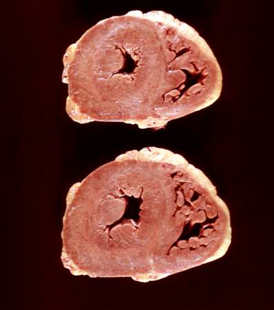
| 19:59, 20 August 2013 | IPLab6Scleroderma5.jpg (file) |  |
19 KB | This is a gross photograph of the heart from this case. There is thickening of the left ventricular wall and some thickening of the right ventricle as well. | 1 |
| 15:20, 20 August 2013 | IPLab5Gout8.jpg (file) |  |
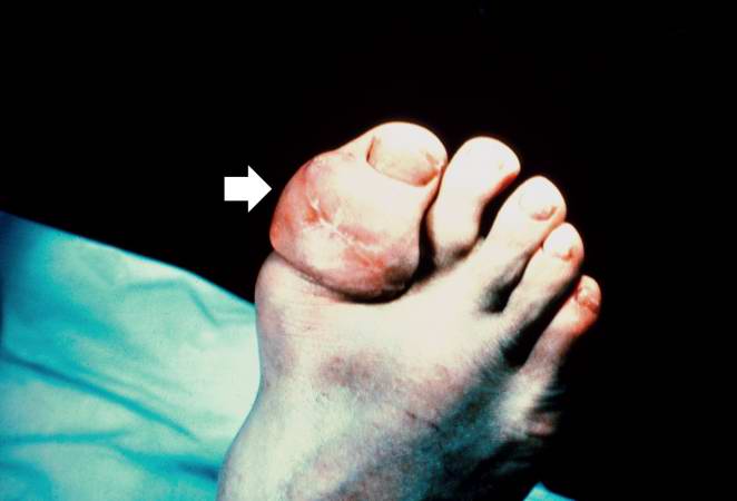
20 KB | This is a gross photograph of a tophus on the great toe of another patient with gout (arrow). The healed surgical incision and the size of this tophus indicate that this was a long-standing problem for this patient. | 1 |
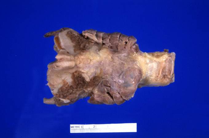
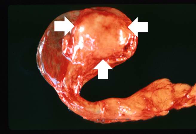
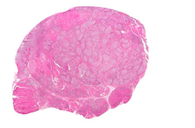
| 17:42, 20 August 2013 | IPLab6Hashimoto1.jpg (file) | 20 KB | This is a gross photograph of thyroid gland taken at autopsy. The gland is only slightly enlarged and has a firm texture. | 1 | |
| 15:18, 20 August 2013 | IPLab5Gout3.jpg (file) |  |
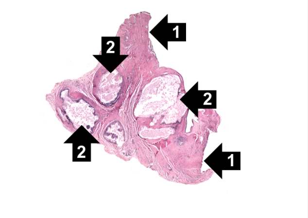
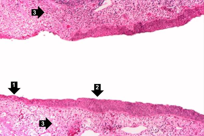
21 KB | This is a low-power photomicrograph of the tophus removed from the elbow of this patient. Note the fibrous connective tissue (1) and the large foci containing the urate crystals (2) surrounded by the intense chronic inflammatory reaction. | 1 |
| 15:17, 20 August 2013 | IPLab5Gout1.jpg (file) |  |
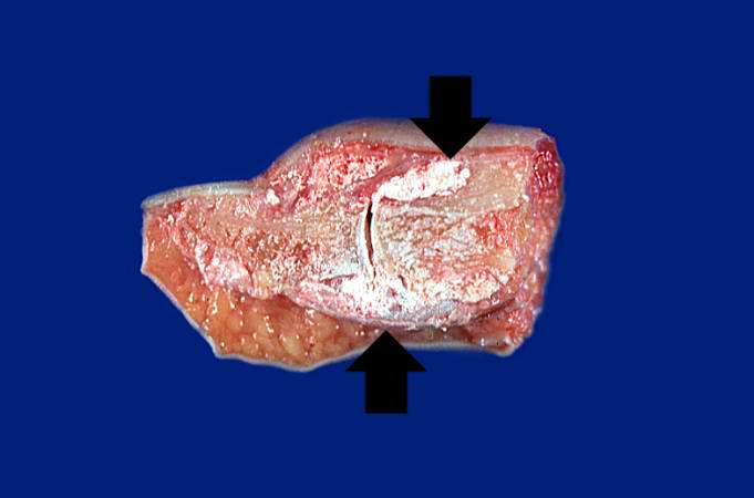
22 KB | This is a gross photograph of an index finger from a patient with gout. The finger has been sectioned longitudinally to demonstrate the distal interphalangeal joint. Note the white chalky material within and adjacent to the joint (arrows). | 1 |
| 16:54, 19 August 2013 | IPLab2FattyChange2.jpg (file) |  |
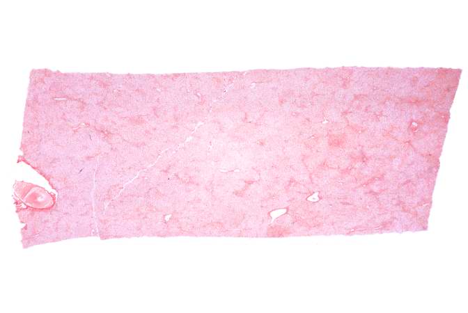
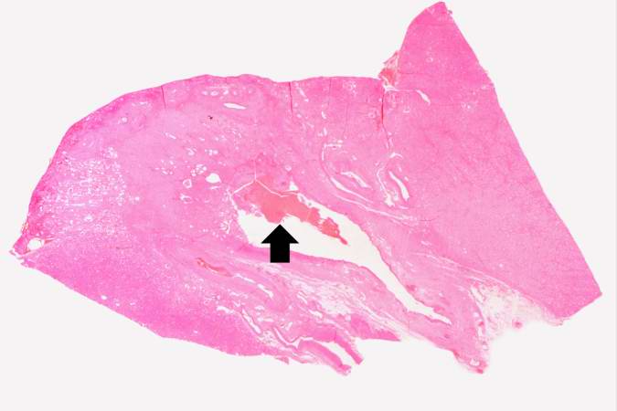
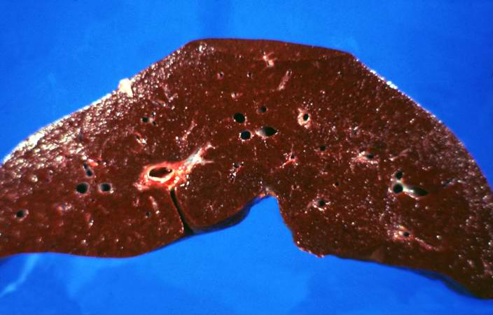
22 KB | This low-power photomicrograph of liver illustrates a very pale-staining section with a uniform appearance throughout the section. | 1 |
| 16:00, 19 August 2013 | IPLab2Atrophy1.jpg (file) |  |
23 KB | This is a gross photograph of an atrophied testis (arrows). | 1 |
| 14:53, 20 August 2013 | IPLab5Hemochromatosis3.jpg (file) |  |
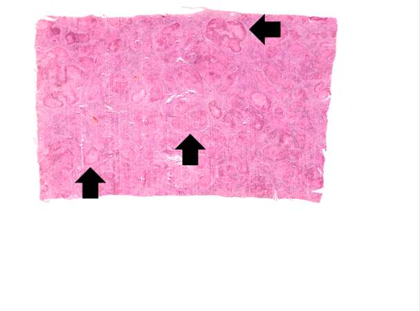
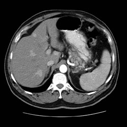
23 KB | This is a low-power micrograph of liver from this patient. Note the nodularity of the tissue (arrows). | 1 |
| 15:10, 20 August 2013 | IPLab5Gaucher3.jpg (file) |  |
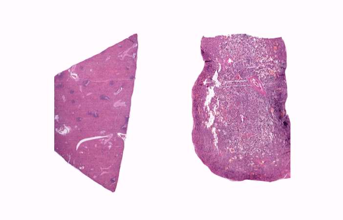
24 KB | This is a low-power photomicrograph of normal spleen (left) and the spleen from this case (right). The loose appearance of the tissue in the Gaucher spleen is due to artifactual loss of tissue during histologic processing. | 1 |
| 14:55, 20 August 2013 | IPLab5Hemochromatosis7.jpg (file) |  |
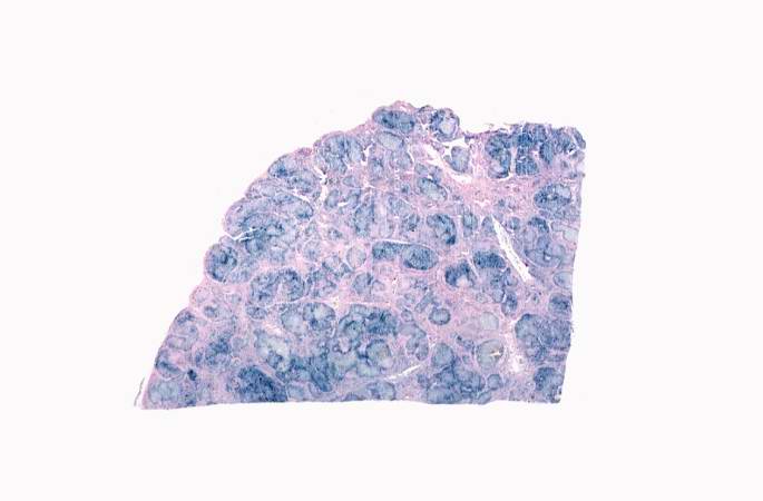
24 KB | This is a low-power view of liver section stained with Prussian blue. Prussian blue reacts with iron in the tissue to give a blue color. | 1 |
| 18:26, 19 August 2013 | IPLab5Antitrypsin7.jpg (file) |  |
25 KB | This is a gross photograph of the cut section of liver from this case. In this view the liver looks smaller than normal and there is a definite micronodular appearance. | 1 |
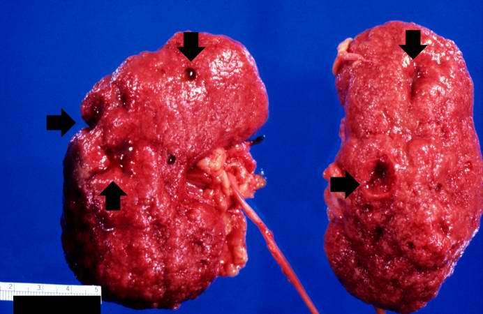
| 17:40, 19 August 2013 | IPLab5PolycysticKidney2.jpg (file) |  |
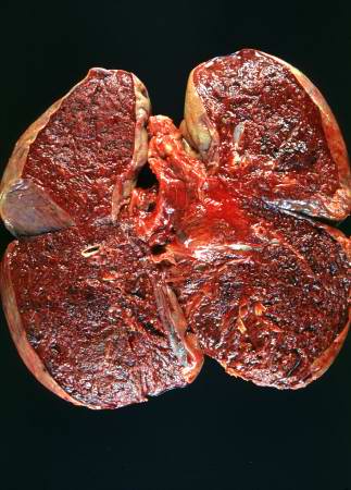
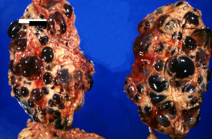
26 KB | This is a gross photograph of the kidneys from this case. Note that both kidneys contain multiple large cysts (arrows). | 1 |
| 15:39, 19 August 2013 | IPLab2Metaplasia1.jpg (file) |  |
26 KB | This is a low-power photomicrograph showing the full cortical and medullary thickness of the kidney. Note that there is a dilated calyx containing some red blood cells in the center of the section (arrow). The cortex is markedly thin and has severe les... | 1 |
| 15:18, 20 August 2013 | IPLab5Gout2.jpg (file) |  |
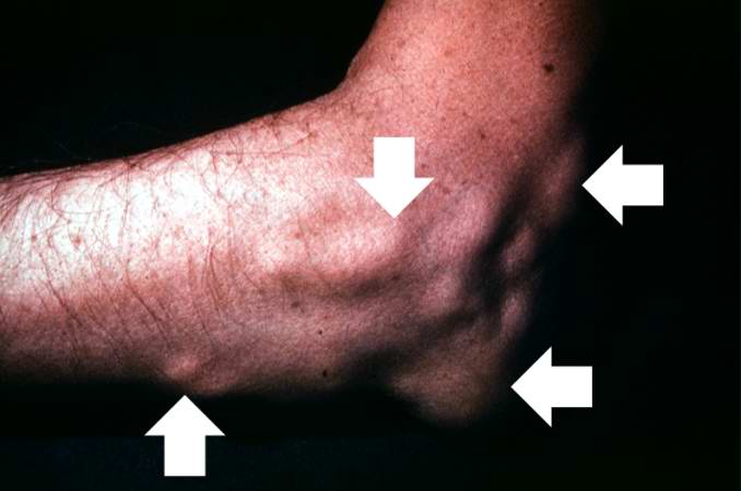
26 KB | This is a gross photograph of the elbow of this patient. The subcutaneous nodules (arrows) on this arm are tophi caused by gout. | 1 |
| 17:42, 20 August 2013 | IPLab6Hashimoto2.jpg (file) | 27 KB | This is a low-power photomicrograph of thyroid from this case. Note that the tissue is more cellular than one would expect and there does not appear to be normal colloid-filled blue spaces in this gland. | 1 | |
| 20:30, 20 August 2013 | IPLab6GN10.jpg (file) |  |
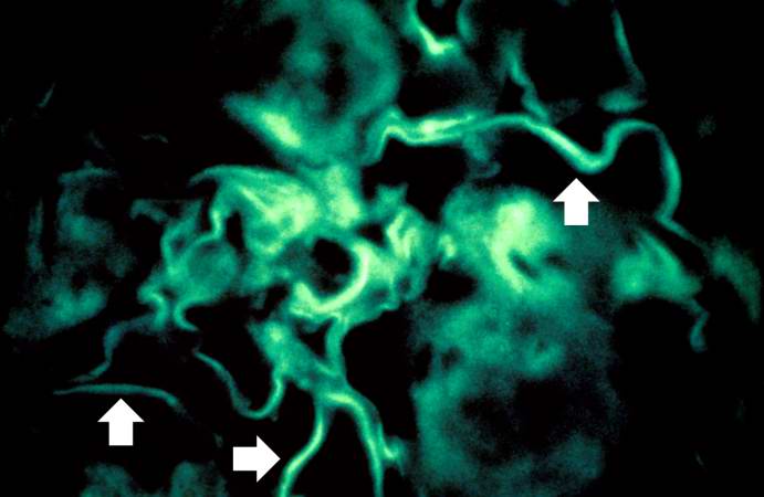
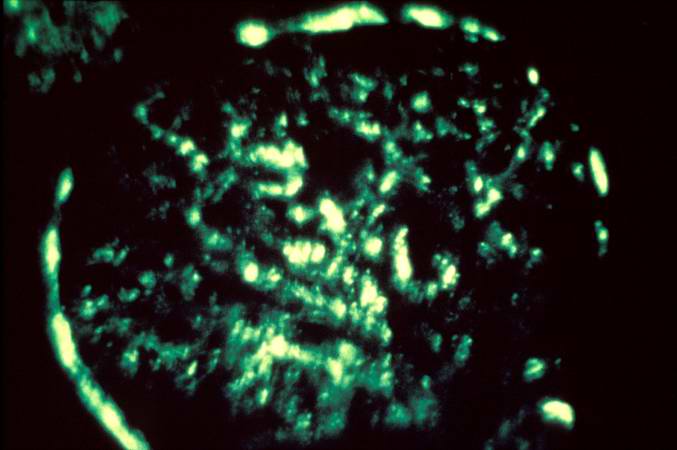
29 KB | For comparison this is an immunofluorescent photomicrograph of a glomerulus from a patient with Goodpasture's syndrome. The linear (arrows) immunofluorescence is characteristic of Goodpasture's syndrome. | 1 |
| 18:25, 19 August 2013 | IPLab5Antitrypsin6.jpg (file) |  |
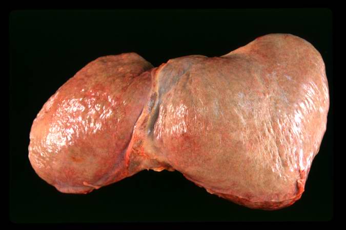
29 KB | This is a gross photograph of the liver from this case. The capsule is somewhat thickened and the surface is slightly roughened, though it is difficult to appreciate the nodularity of the liver. | 1 |
| 01:50, 9 July 2020 | IPLab7Metastatic1a.jpg (file) |  |
30 KB | 1 | |
| 20:28, 20 August 2013 | IPLab6GN5.jpg (file) |  |
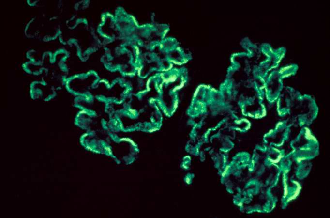
30 KB | This is an immunofluorescent photomicrograph of granular membranous immunofluorescence (immune complex disease). The antibody used for these studies was specific for IgG. | 1 |
| 20:30, 20 August 2013 | IPLab6GN8.jpg (file) |  |
31 KB | This immunofluorescent photomicrograph of a glomerulus from a case of acute poststreptococcal glomerulonephritis shows a granular immunofluorescence pattern consistent with immune complex disease. The primary antibody used for this staining was specifi... | 1 |
| 17:19, 19 August 2013 | IPLab5Neurofibromatosis3.jpg (file) |  |
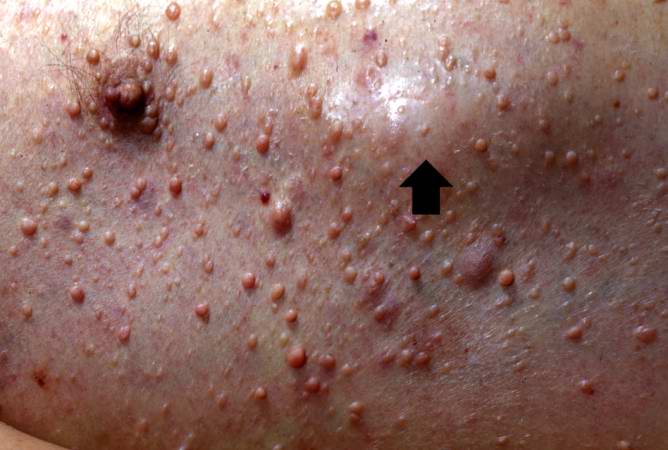
32 KB | This is a closer view of neurofibromas on the skin. | 1 |
| 18:07, 19 August 2013 | IPLab5Antitrypsin2.jpg (file) |  |
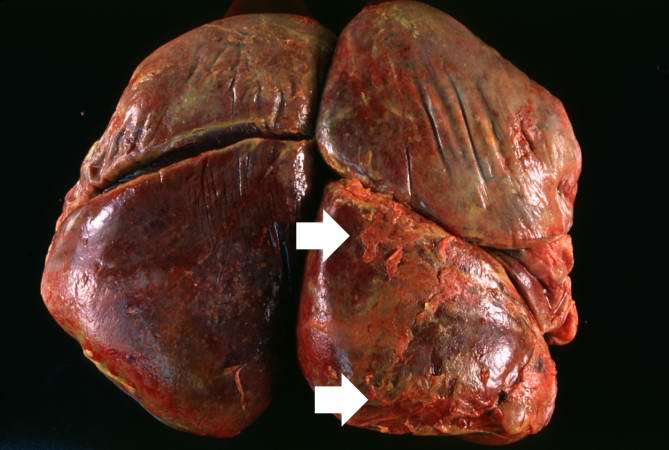
32 KB | This is a gross photograph of the cut sections of lung from this case. The lung parenchyma is markedly hemorrhagic and consolidated. Again the hemorrhage makes it difficult to appreciate the emphysematous changes. | 1 |
| 17:49, 19 August 2013 | IPLab5PolycysticKidney3.jpg (file) |  |
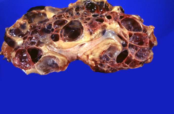
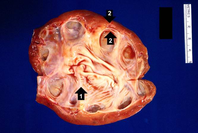
33 KB | This is a gross photograph of a cut section from one of these polycystic kidneys. Note that the renal parenchyma is almost completely replaced by cystic structures. | 1 |
| 18:26, 19 August 2013 | IPLab5Antitrypsin8.jpg (file) |  |
35 KB | This is a closer view of the cut section of liver from this case. There is a definite micronodular pattern to the liver parenchyma. | 1 |
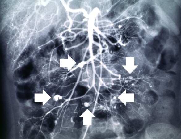
| 17:56, 20 August 2013 | IPLab6PAN3.jpg (file) |  |
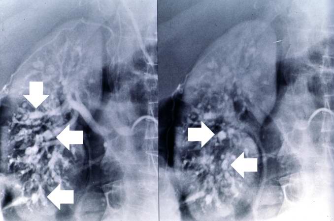
36 KB | This angiogram of the kidneys demonstrates numerous aneurysmal dilatations in the renal circulation (arrows). | 1 |
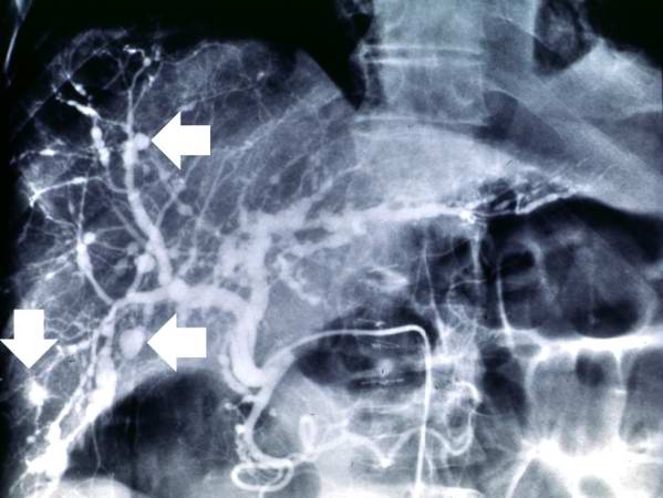
| 17:55, 20 August 2013 | IPLab6PAN1.jpg (file) |  |
36 KB | This angiogram of the abdominal viscera demonstrates numerous aneurysms throughout the mesenteric circulation (arrows). | 1 |
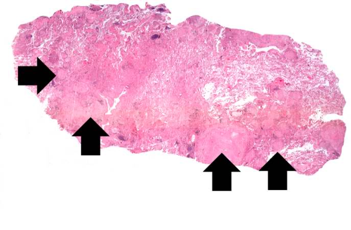
| 20:10, 20 August 2013 | IPLab6TB2.jpg (file) |  |
36 KB | This is a low-power photomicrograph of lung tissue with multiple circumscribed nodules - granulomas (arrows). | 1 |
| 17:55, 20 August 2013 | IPLab6PAN2.jpg (file) |  |
36 KB | This angiogram of the liver also demonstrates numerous aneurysms throughout the hepatic circulation (arrows). | 1 |
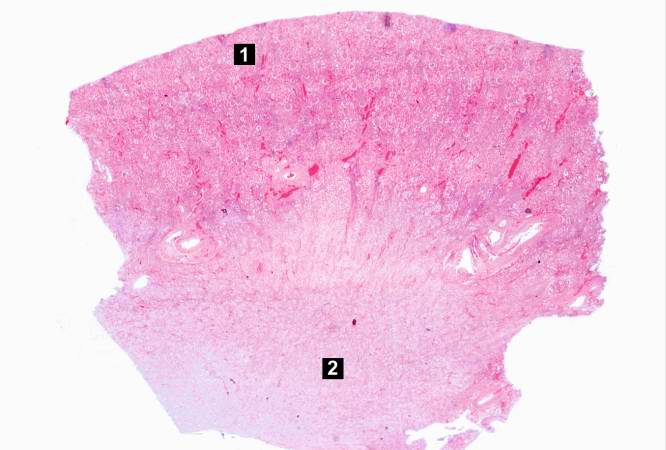
| 15:26, 20 August 2013 | IPLab5DM2.jpg (file) |  |
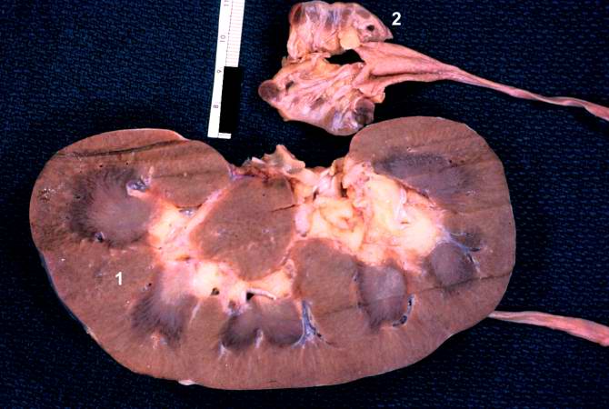
36 KB | This is a low-power photomicrograph of the kidney from this patient. The section extends from cortex (1) to the medulla (2). | 1 |
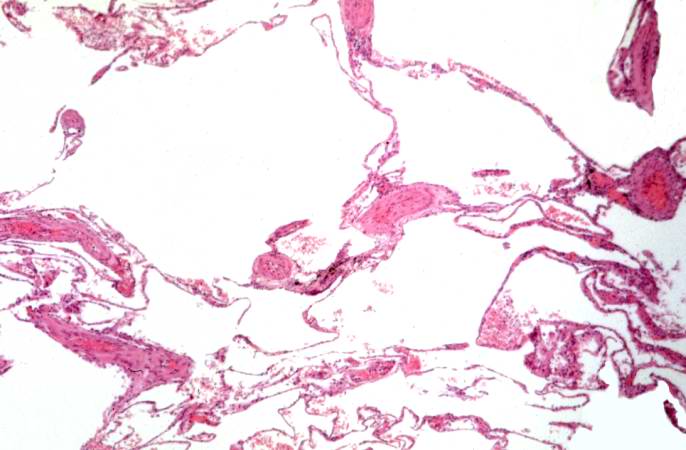
| 16:38, 19 August 2013 | IPLab2Calcification2.jpg (file) |  |
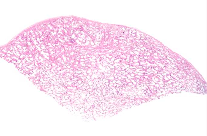
37 KB | This low-power photomicrograph of the patient's lung illustrates large, open alveolar spaces. The pleural surface is the curved surface at the top. | 1 |
| 20:14, 20 August 2013 | IPLab6TB6.jpg (file) |  |
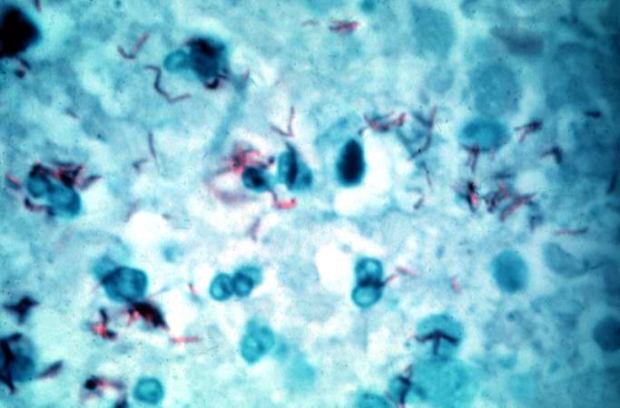
37 KB | This is a high-power (oil immersion) photomicrograph of granuloma stained with an acid-fast stain. Mycobacterium tuberculosis bacilli stain red. | 1 |
| 18:06, 19 August 2013 | IPLab5Antitrypsin1.jpg (file) |  |
37 KB | This is a gross photograph of the lungs from this case. The rough friable material on the surface of the lung (arrows) is fibrinous exudate and fibrous tissue. This reaction on the surface of the lung is due to the recent surgery. The emphysematous cha... | 1 |
| 15:29, 19 August 2013 | IPLab2Hyperplasia9.jpg (file) |  |
37 KB | This kidney was removed from another autopsy patient who had prostatic hyperplasia resulting in marked urinary retention and back-flow of urine from the bladder into the ureters and renal pelvis. The increased pressure inside the renal pelvis resulted ... | 1 |
| 14:53, 20 August 2013 | IPLab5Hemochromatosis2.jpg (file) |  |
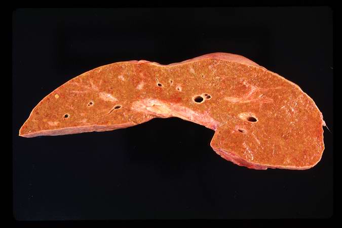
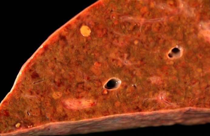
37 KB | This is a gross photograph of a cut section of liver from this case of hemochromatosis. Note that the liver is dark brown. Although hard to appreciate in a photograph, the tissue is also firm (cirrhotic). | 1 |
| 16:35, 19 August 2013 | IPLab2Calcification6.jpg (file) |  |
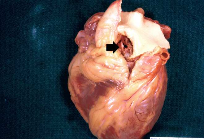
39 KB | Metastatic calcification is only one of two forms of pathologic calcification. Unlike metastatic calcification, dystrophic calcification does not require an increase in serum calcium levels. This is a gross specimen of a heart with dystrophic calcifica... | 1 |
| 17:19, 19 August 2013 | IPLab5Neurofibromatosis2.jpg (file) |  |
40 KB | This is another view taken at autopsy demonstrating the neurofibromas. Some lesions can be seen as subcutaneous swellings (arrow) and others form pedunculated masses. Most are hyperpigmented. | 1 |
| 15:40, 19 August 2013 | IPLab2Metaplasia2.jpg (file) |  |
41 KB | his high-power photomicrograph demonstrates the transitional epithelium lining the renal calyx (1) and the junction (transition zone) to a thicker hyperplastic epithelium (2). Note the inflammatory cells and increased vascular response in the stromal t... | 1 |
| 18:09, 19 August 2013 | IPLab5Antitrypsin5.jpg (file) |  |
41 KB | This is a low-power photomicrograph from an area of the lung without significant hemorrhage. The enlarged, emphysematous air spaces are easily appreciated. | 1 |
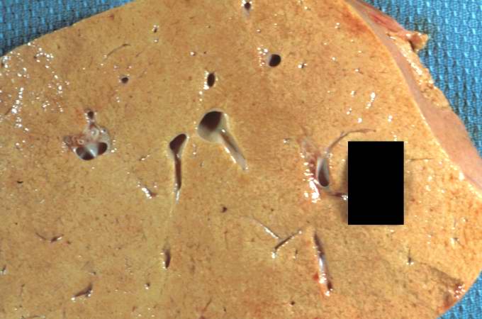
| 16:53, 19 August 2013 | IPLab2FattyChange1.jpg (file) |  |
42 KB | This gross photograph of liver tissue illustrates the yellowish color of the liver parenchyma. The yellow color indicates high fat content in this tissue. Compare this with the normal dark red color of liver. | 1 |
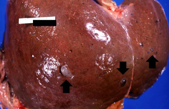
| 17:53, 19 August 2013 | IPLab5PolycysticKidney8.jpg (file) |  |
42 KB | This is a gross photograph of the liver from this patient. Multiple cysts can be seen on the surface of this liver (arrows). | 1 |
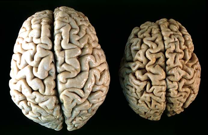
| 16:12, 19 August 2013 | IPLab2Atrophy10.jpg (file) |  |
43 KB | This gross photograph shows a normal brain (left) and a brain from a geriatric patient (right). Note the decreased size, the narrowed gyri, and the widened sulci of the brain from this octogenarian. What is the cause of atrophy in this case? | 1 |
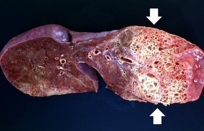
| 19:58, 20 August 2013 | IPLab6Scleroderma2.jpg (file) |  |
43 KB | This is a gross photograph of a cut section of one lung from this patient. Note the extensive fibrosis lower lobe (arrows). | 1 |
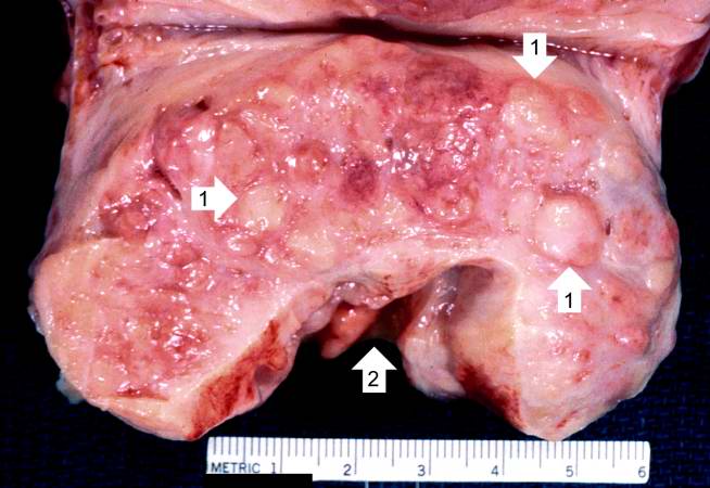
| 15:27, 19 August 2013 | IPLab2Hyperplasia2.jpg (file) |  |
44 KB | This is a close-up of the prostate from this same patient. Note the nodularity of the tissue (1) and the enlargement of the gland. Enlargement of the prostate leads to compression of the urethra as it passes through (2) the gland. | 1 |
| 15:26, 20 August 2013 | IPLab5DM1.jpg (file) |  |
44 KB | This is a gross photograph of the kidneys from this case. Note that there are multiple shrunken regions (old infarcts) (arrows) and the kidneys have a rough granular appearance on the surface, which is caused by multiple small infarcts of small vessels... | 1 |
| 16:36, 19 August 2013 | IPLab2Calcification8.jpg (file) |  |
45 KB | This gross photograph affords a closer view of the same aortic valve. Note the nodularity and thickening of this valve due to fibrosis and dystrophic calcification. | 1 |
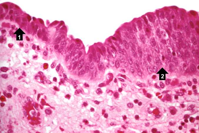
| 15:43, 19 August 2013 | IPLab2Metaplasia4.jpg (file) |  |
46 KB | This is a higher-power photomicrograph of the junction of normal epithelium (1) with hyperplastic transitional epithelium (2). | 1 |
| 16:11, 19 August 2013 | IPLab2Atrophy9.jpg (file) |  |
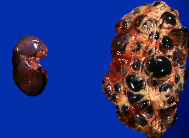
47 KB | The two kidneys in this slide are from the same patient. One kidney (1) is relatively normal, although increased in size due to compensatory hypertrophy. The other kidney (2) is very small with only rudimentary nodules of renal parenchyma. This kidney ... | 1 |
| 17:39, 19 August 2013 | IPLab5PolycysticKidney1.jpg (file) |  |
48 KB | This is a gross photograph of the kidneys from this case. Note that both kidneys contain multiple large cysts (arrows). | 1 |
| 15:28, 19 August 2013 | IPLab2Hyperplasia3.jpg (file) |  |
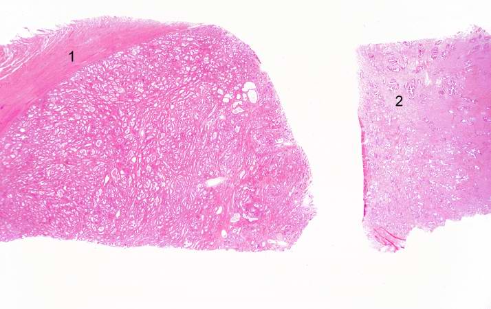
48 KB | This is a low-power photomicrograph showing hyperplastic prostate on the left (1) and normal prostate on the right (2). At this power, dilated glands are visible in the section of hyperplastic prostate. | 1 |
| 17:19, 19 August 2013 | IPLab5Neurofibromatosis4.jpg (file) |  |
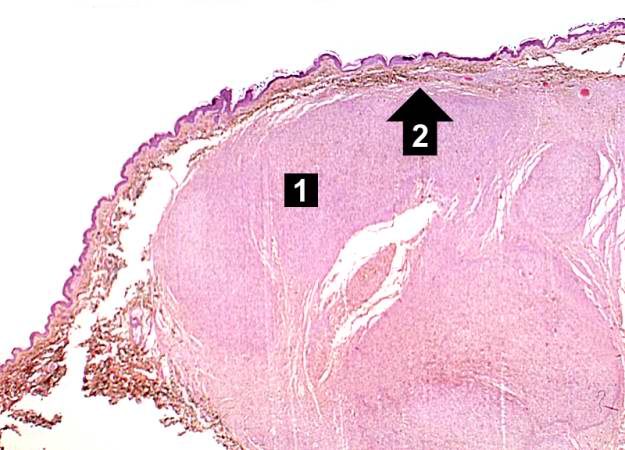
48 KB | This is a low-power photomicrograph of a subcutaneous neurofibroma (1). Note the increased pigmentation in the skin (2). | 1 |