File list
This special page shows all uploaded files.
| Date | Name | Thumbnail | Size | Description | Versions |
|---|---|---|---|---|---|
| 14:52, 8 August 2013 | Test.mp3 (file) | 5.65 MB | Testing out Html5mediator | 1 | |
| 17:19, 8 August 2013 | Test.mp4 (file) | 4.17 MB | More testing! | 2 | |
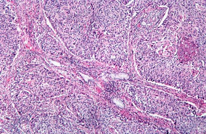
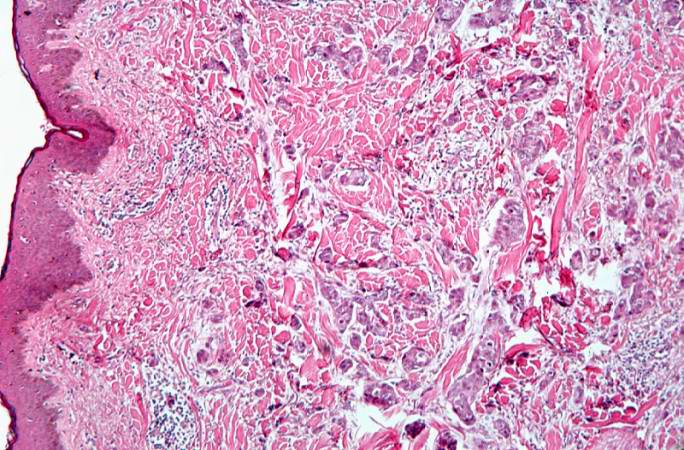
| 20:29, 16 January 2014 | CytologicallyYoursUnknowns201401-04-10.jpg (file) |  |
730 KB | 3 | |
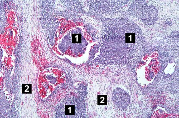
| 20:28, 16 January 2014 | CytologicallyYoursUnknowns201401-04-08.jpg (file) |  |
679 KB | Reverted to version as of 20:27, 16 January 2014 | 4 |
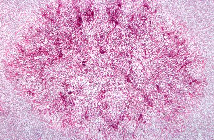
| 20:29, 16 January 2014 | CytologicallyYoursUnknowns201401-04-09.jpg (file) |  |
613 KB | 2 | |
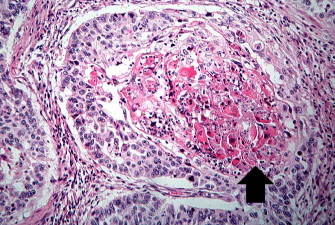
| 20:27, 16 January 2014 | CytologicallyYoursUnknowns201401-04-07.jpg (file) |  |
525 KB | 2 | |
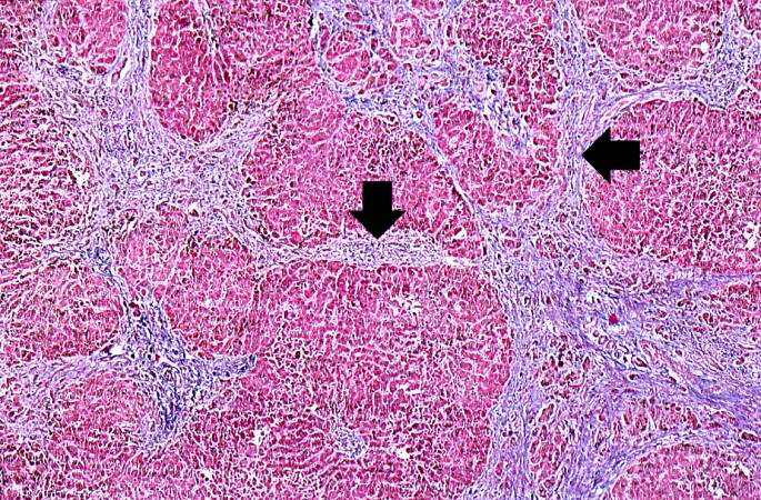
| 05:13, 21 August 2013 | IPLab12Alcoholic4.jpg (file) |  |
130 KB | In this is medium-power photomicrograph of trichrome stained liver the bands of fibrous tissue are seen to form "bridges" between triad areas (arrows); this is called "bridging fibrosis." Also note the fibrous tissue (arrows) and how the hepatocytes ar... | 1 |
| 05:14, 21 August 2013 | IPLab12Alcoholic5.jpg (file) |  |
122 KB | In this high-power photomicrograph of trichrome-stained liver, the bands of fibrous tissue surround the hepatocyte nodules. There is some degeneration and dropout of hepatocytes in this nodule. Also note the increased numbers of bile ducts in the triad... | 1 |
| 05:33, 21 August 2013 | IPLab12Mesothelioma4.jpg (file) |  |
113 KB | This is a low-power photomicrograph of a section of the left lung. At this magnification you can see areas of consolidation (arrows), especially around blood vessels. | 1 |
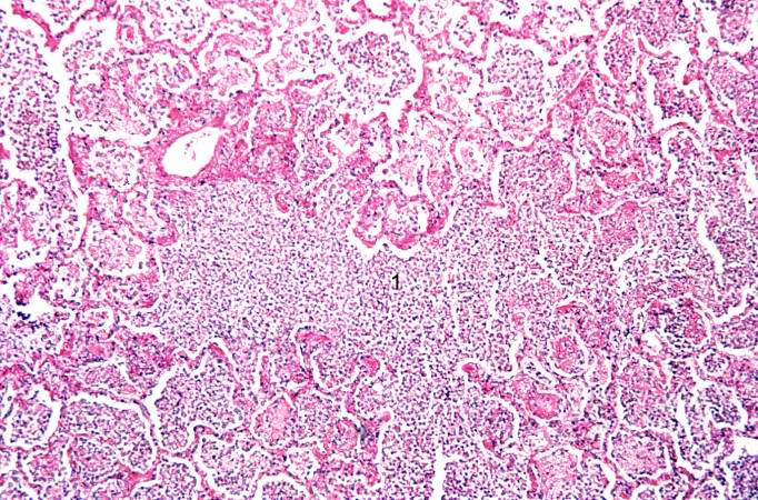
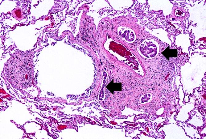
| 03:23, 19 August 2013 | IPLab3Bronchopneumonia5.jpg (file) |  |
113 KB | This is a photomicrograph of lung tissue affected by bronchopneumonia. Note that the alveolar structure of this tissue, which is in the region of a terminal bronchiole (1), has been retained despite the massive infiltration of inflammatory cells. These... | 1 |
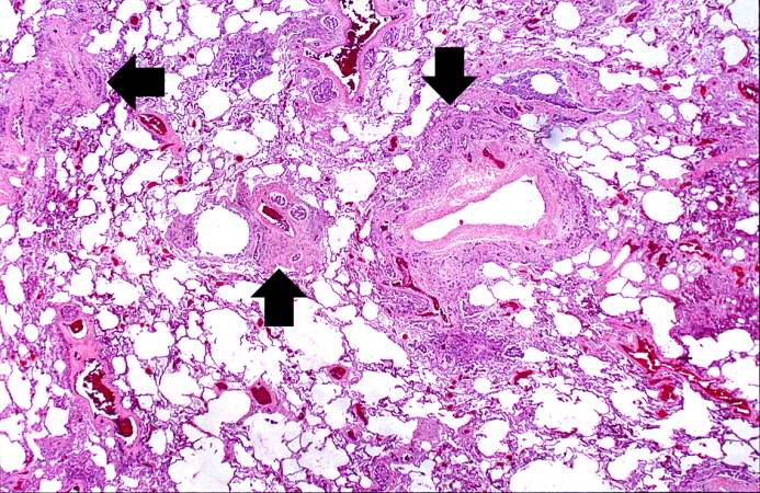
| 05:25, 21 August 2013 | IPLab12RadiationFibrosis9.jpg (file) |  |
107 KB | This is a photomicrograph of an area of tissue exhibiting diffuse fibrosis and thickening of the alveolar septa. | 1 |
| 02:12, 19 August 2013 | IPLab3AcuteAppendicitis6.jpg (file) |  |
106 KB | This higher-power photomicrograph of the mucosal surface shows the loss of normal mucosal epithelium (arrows) and the inflammatory infiltrate. The principal inflammatory cell in this case of acute appendicitis is the neutrophil. | 1 |
| 05:16, 21 August 2013 | IPLab12Alcoholic10.jpg (file) |  |
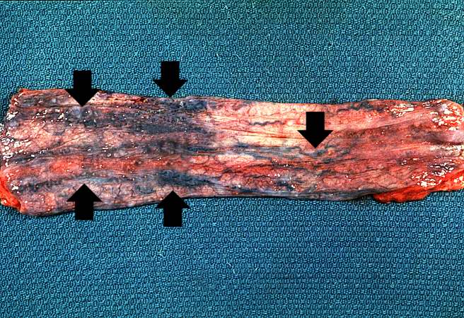
105 KB | This is a photograph taken from another patient at autopsy to demonstrate numerous esophageal varices in the distal esophagus (arrows). None of these varices have ruptured. | 1 |
| 04:15, 19 August 2013 | IPLab3ChronicPepticUlcer4.jpg (file) |  |
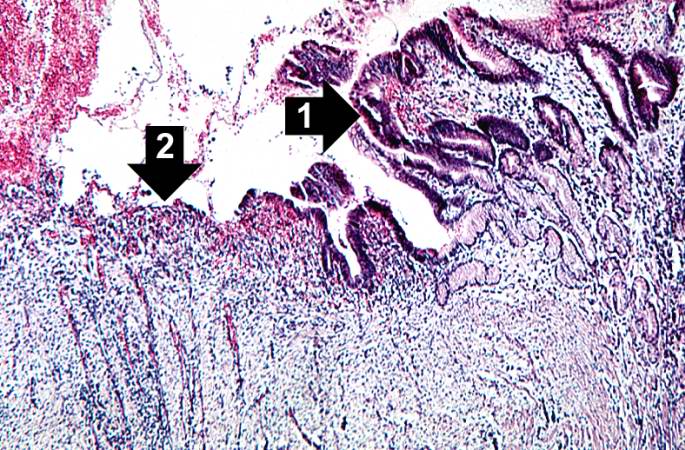
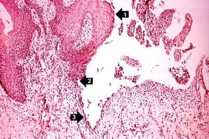
104 KB | This is a photomicrograph of the margin of the ulcer. Note the intact epithelium on the right side of the section (1) and the ulcerated region without epithelium on the left (2). There are numerous inflammatory cells within this tissue. | 1 |
| 04:16, 21 August 2013 | IPLab10Blasto6.jpg (file) |  |
103 KB | This high-power photomicrograph shows what appear to be inflammatory cells filling the alveoli. At this magnification, numerous round bodies (arrows) that look like inflammatory cell nuclei can be seen. However, on closer examination, some of these rou... | 1 |
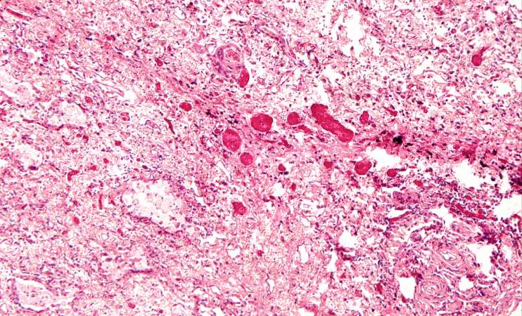
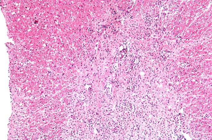
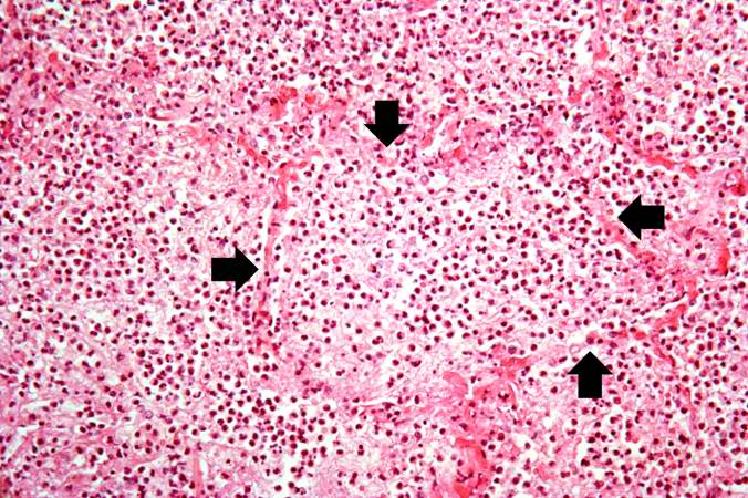
| 05:19, 21 August 2013 | IPLab12Acetaminophen2.jpg (file) |  |
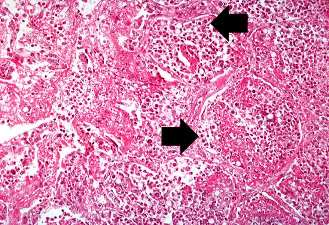
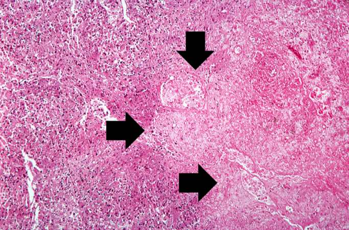
103 KB | In this photomicrograph of the liver from this patient there are areas of hemorrhagic necrosis (arrows). | 1 |
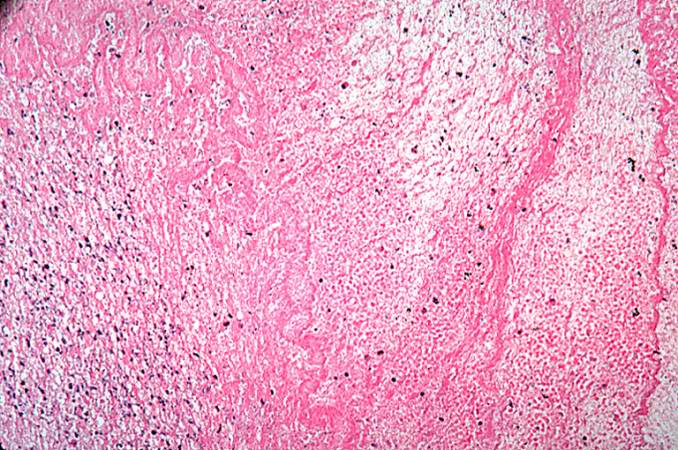
| 05:13, 21 August 2013 | IPLab12Alcoholic3.jpg (file) |  |
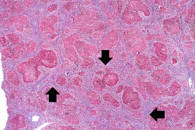
102 KB | This is a low-power photomicrograph of this liver stained with a trichrome stain to highlight the fibrous tissue (arrows). Also note the nodular pattern. | 1 |
| 01:33, 21 August 2013 | IPLab7LipSCC8.jpg (file) |  |
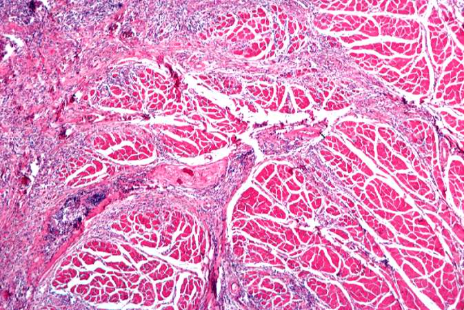
101 KB | This is a section of muscle tissue from this biopsy of the lip. Note that the squamous cell carcinoma has infiltrated into the muscle tissue. There are also inflammatory cells within this area of tumor infiltration. | 1 |
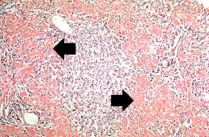
| 05:19, 21 August 2013 | IPLab12Acetaminophen3.jpg (file) |  |
100 KB | This is another example of the areas of hemorrhagic necrosis (arrows). | 1 |
| 05:33, 21 August 2013 | IPLab12Mesothelioma7.jpg (file) |  |
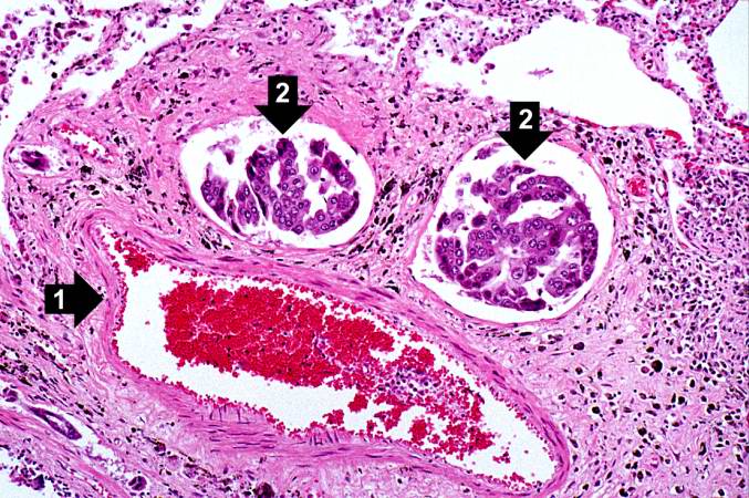
100 KB | In this higher-power photomicrograph there is a blood vessel (1) and adjacent lymphatics that contain tumor cells (2). There are also accumulations of brown material adjacent to these vessels. | 1 |
| 05:33, 21 August 2013 | IPLab12Mesothelioma5.jpg (file) |  |
99 KB | This higher-power photomicrograph of left lung shows areas of consolidation and fibrosis (arrows). Also note that in many of these areas there are clusters of blue cells. | 1 |
| 02:00, 21 August 2013 | IPLab7Bronchogenic4.jpg (file) |  |
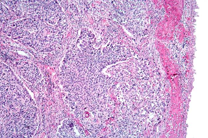
99 KB | This is a higher-power photomicrograph of bronchus with the ulcerated mucosal surface on the right and tumor underneath. | 1 |
| 02:00, 21 August 2013 | IPLab7Bronchogenic6.jpg (file) |  |
98 KB | This is a photomicrograph of tumor from an area of invasion with compression of fibrous stroma and focal necrosis. | 1 |
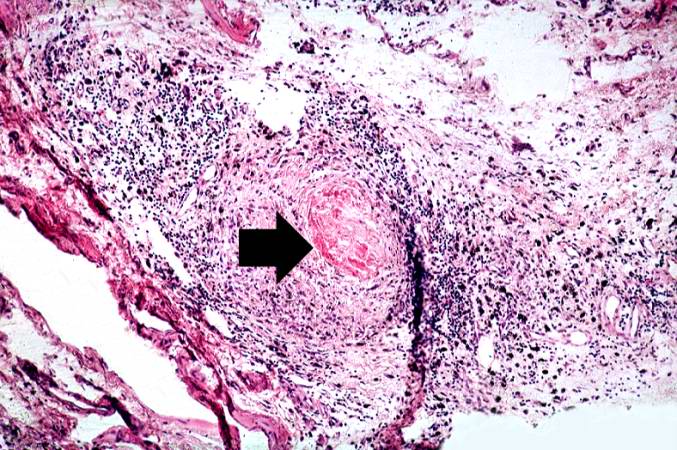
| 18:06, 19 August 2013 | IPLab6RA2.jpg (file) |  |
97 KB | This is a medium-power photomicrograph of the joint capsule surrounding the metacarpal joints. Note the thickening of the capsule and the focal accumulation of inflammatory cells surrounding a central area of fibrinoid necrosis (arrow). | 1 |
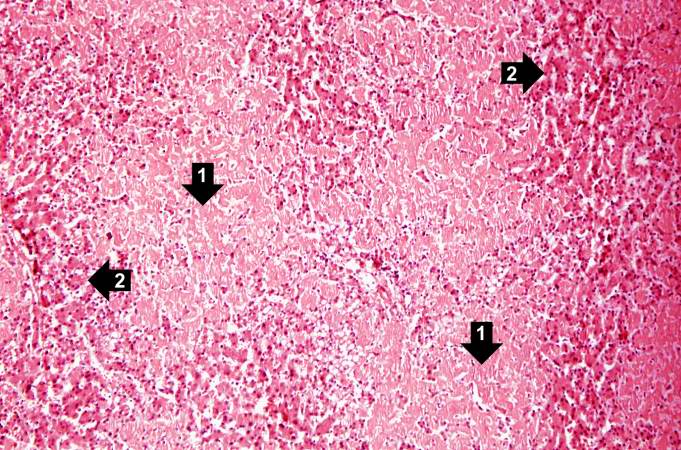
| 21:34, 20 August 2013 | IPLab6Amyloid4.jpg (file) |  |
97 KB | This is a low-power photomicrograph of liver tissue from this case. Note the eosinophilic hyaline material (1) present within and between the hepatic tissue (2). There is marked distortion of lobular architecture by the amyloid. | 1 |
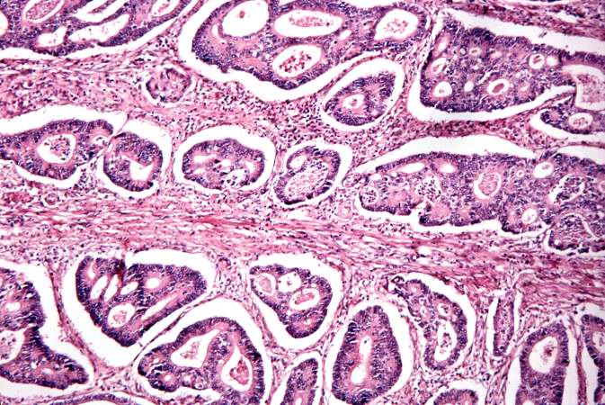
| 01:41, 21 August 2013 | IPLab7ColonCA6.jpg (file) |  |
96 KB | This is a high-power photomicrograph of tumor cells forming glands. | 1 |
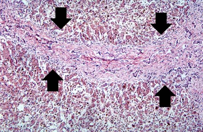
| 05:47, 21 August 2013 | IPLab13BiliaryAtresia2.jpg (file) |  |
95 KB | This medium-power photomicrograph of liver shows an area of portal fibrosis and bile duct proliferation (arrows). Adjacent to this fibrotic portal region, hepatocytes are seen separated by dilated sinusoids. Throughout this section are found accumulati... | 1 |
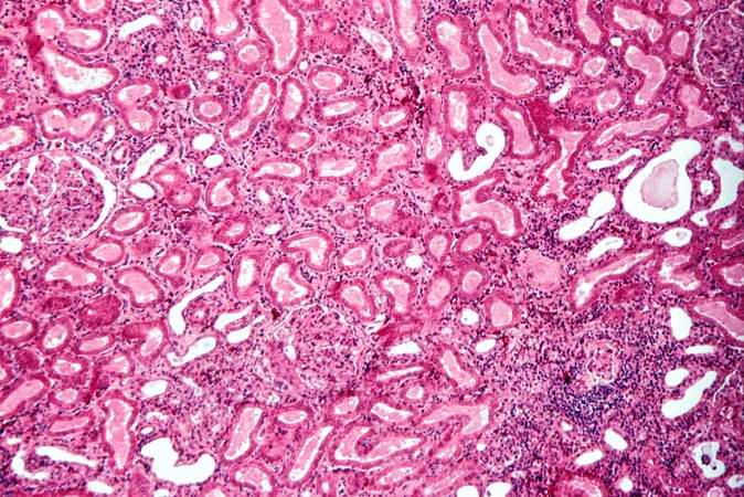
| 21:57, 20 August 2013 | IPLab6AcuteRejection6.jpg (file) |  |
95 KB | This is a higher-power photomicrograph demonstrating the cellular infiltrate within the interstitium. There is some degeneration (coagulative necrosis) of tubules and glomeruli. | 1 |
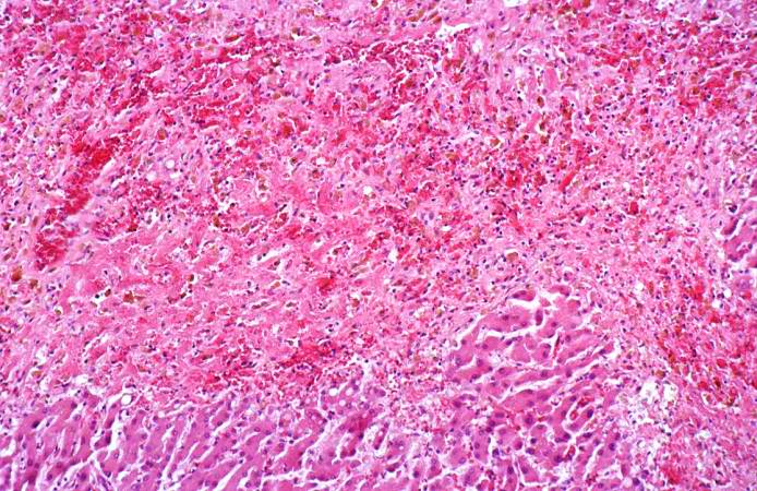
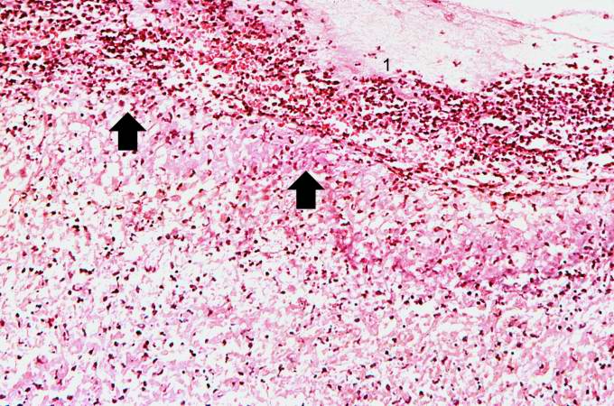
| 05:19, 21 August 2013 | IPLab12Acetaminophen4.jpg (file) |  |
95 KB | This is a medium-power photomicrograph of the areas of hemorrhagic necrosis. Note the coagulation necrosis and hemorrhage in this area. Viable hepatocytes can be seen along the edge of this lesion. | 1 |
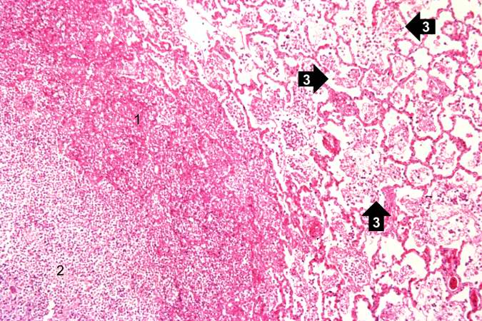
| 03:23, 19 August 2013 | IPLab3Bronchopneumonia4.jpg (file) |  |
94 KB | This photomicrograph of the wall of an abscess (1) illustrates liquefaction necrosis in the center of the abscess (2). The remaining lung parenchyma (on the right side of the image) has extensive neutrophilic infiltration into the alveoli (3). This abs... | 1 |
| 02:43, 21 August 2013 | IPLab8Polio3.jpg (file) |  |
93 KB | This is a high-power photomicrograph of the anterior horn of the spinal cord from this case. Note the absence of motor neurons and the diffuse gliosis. | 1 |
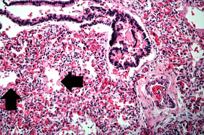
| 05:51, 21 August 2013 | IPLab13Hyaline7.jpg (file) |  |
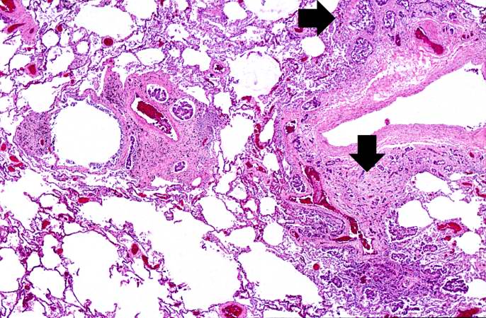
93 KB | This high-power photomicrograph shows an airway with adjacent lung tissue. Some alveoli have hyaline membranes (arrows). There is severe congestion of the interstitium throughout this section. | 1 |
| 04:16, 19 August 2013 | IPLab3ChronicPepticUlcer5.jpg (file) |  |
93 KB | This is a medium-power photomicrograph of the base of the ulcer with the fibrinopurulent membrane (1) overlying the ulcerated surface. The ulcerated surface contains granulation tissue and inflammatory cells (2). | 1 |
| 16:29, 19 August 2013 | IPLab4MuralThrombus5.jpg (file) |  |
93 KB | This photomicrograph illustrates the layered effect of the thrombus. | 1 |
| 01:56, 21 August 2013 | IPLab7Melanoma5.jpg (file) |  |
93 KB | This is a higher-magnification showing the abundant extracellular melanin (arrows) surrounding the tumor cells. This section of neoplasm shows the numerous cells with abundant cytoplasm and brown pigment within the cytoplasm of some of these cells. | 1 |
| 03:18, 19 August 2013 | IPLab3LobarPneumonia8.jpg (file) |  |
92 KB | This is a photomicrograph of alveoli filled with exudate. The alveolar wall outlines (arrows) are barely visible in this section. The alveoli are filled with PMNs, fibrin, and edema fluid. This is a severe acute inflammatory response but the structure ... | 1 |
| 04:16, 19 August 2013 | IPLab3ChronicPepticUlcer6.jpg (file) |  |
92 KB | This high-power photomicrograph of the ulcer base (arrows) demonstrates the lack of epithelium and the exuberant inflammatory response (1) consisting of primarily of fibrin (and adherent gastric secretions) and PMNs. The surface of the ulcer bed is cov... | 1 |
| 21:35, 20 August 2013 | IPLab6Amyloid6.jpg (file) |  |
92 KB | This is a low-power photomicrograph of liver tissue stained with Congo red (orange color in slide). Congo red reacts with amyloid and gives it an orange color (arrows). | 1 |
| 05:33, 21 August 2013 | IPLab12Mesothelioma6.jpg (file) |  |
92 KB | In this higher-power photomicrograph the clusters of tumor cells (arrows) can be appreciated. | 1 |
| 01:50, 21 August 2013 | IPLab7IDC3.jpg (file) |  |
91 KB | This is a section of breast with small groups of carcinoma cells throughout the breast tissue and invading through the dermis. | 1 |
| 04:03, 21 August 2013 | IPLab10Candidiasis5.jpg (file) |  |
91 KB | This higher-power photomicrograph shows the yeasts and pseudohyphae in this focus of Candida organisms. | 1 |
| 05:55, 21 August 2013 | IPLab13WT5.jpg (file) |  |
91 KB | This low-power photomicrograph of tumor shows the two cell types making up this neoplasm. The basophilic cellular component termed "blastema" (1) can be distinguished from less cellular eosinophilic areas with fibroblast-like cells (2). | 1 |
| 02:01, 21 August 2013 | IPLab7Bronchogenic8.jpg (file) |  |
91 KB | This is a high power photomicrograph of tumor with an area of central necrosis (arrow). | 1 |
| 02:28, 21 August 2013 | IPLab8HSVGlossitis2.jpg (file) |  |
90 KB | This higher-power photomicrograph shows the epithelium (1), the edge of the ulcer (2), and the ulcerated epithelium (3). There is an inflammatory exudate at the base of the ulcer and some necrotic cells where the epithelium once was present. | 1 |
| 04:06, 21 August 2013 | IPLab10Histo3.jpg (file) |  |
90 KB | This is an even higher-power photomicrograph of an area of necrosis (arrows). There is loss of cellular detail within this area. There are inflammatory cells present; however, it is difficult to differentiate the inflammatory cells from the native lymp... | 1 |
| 04:16, 21 August 2013 | IPLab10Blasto7.jpg (file) |  |
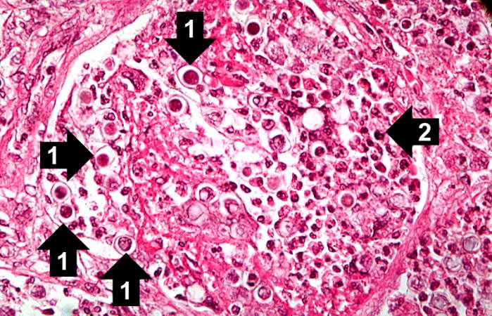
90 KB | This high-power photomicrograph shows an alveolus filled with numerous round bodies up to 25 mm in diameter. Some of these double-contour bodies (1) have a dense center and a clear halo. These are the Blastomyces organisms. The typical B. dermatitides ... | 1 |
| 05:52, 21 August 2013 | IPLab13Hyaline8.jpg (file) |  |
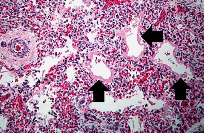
90 KB | This medium-power photomicrograph shows the pink acellular homogeneous material lining the alveoli which comprises the hyaline membranes (arrows). The interstitium shows congestion, as in previous sections. | 1 |
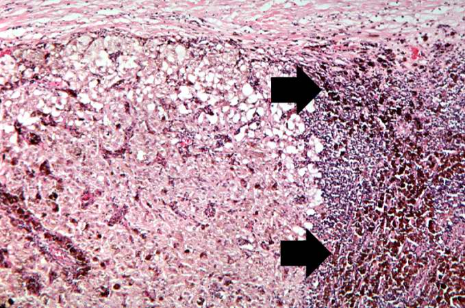
| 21:48, 20 August 2013 | IPLab6ChronicRejection3.jpg (file) |  |
89 KB | This is a photomicrograph of kidney with a focal area of hemorrhage around a small blood vessel (left) and congestion of the glomeruli. Note that there is a marked loss of renal tubules throughout this section with replacement by fibrous connective tis... | 1 |
| 05:51, 21 August 2013 | IPLab13Hyaline3.jpg (file) |  |
89 KB | This is a low-power photomicrograph of liver which contains dark blue-stained cells in the hepatic sinusoids. These are immature blood cell precursors and this represents extramedullary hematopoiesis of the liver. | 1 |
| 03:23, 19 August 2013 | IPLab3Bronchopneumonia6.jpg (file) |  |
89 KB | This is a photomicrograph of another area in the lung showing a terminal bronchiole (1) in which the mucosal lining has been almost completely destroyed. There is extensive neutrophilic infiltration throughout this lung tissue. | 1 |